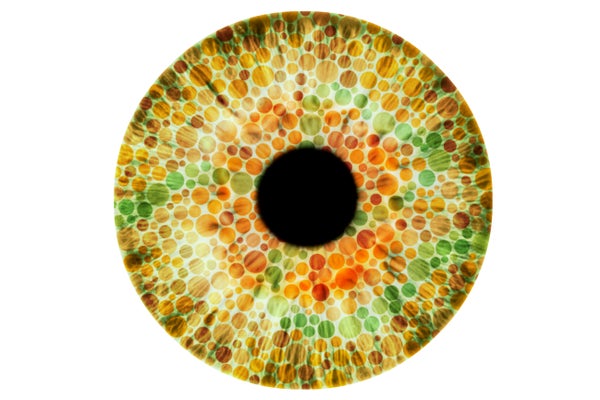
Sight begins when light bounces off surfaces and enters our eyes. The muscles of our pupils control how much light passes through, and the clear cornea and lens bend the light and focus it onto the retina, a thin strip of tissue covered in millions of light-sensitive neurons, or photoreceptors.
These nerve cells, named for the way they are shaped—like rods and cones—are where light is converted into electrical signals then sent via the optic nerve to the visual centers of the brain. A paper published October 11 in Science uses a retina grown outside the body to show how cones develop into the eyes’ color sensors.
Our daytime vision depends on the cones because they respond best to bright light (as opposed to the rods, which are sensitive to dim illumination). The pyramid-shaped cells come in three types: blue, green and red—each named after the colors of light they are able to detect. We need all three to perceive the many hues in our surroundings. The most common cause of color blindness—which affects approximately 8 percent of males and 0.5 percent of females of northern European descent—is caused by an inherited defect in red or green cones, which leads to reduced or complete loss in ability to see the two colors those cells detect.
On supporting science journalism
If you're enjoying this article, consider supporting our award-winning journalism by subscribing. By purchasing a subscription you are helping to ensure the future of impactful stories about the discoveries and ideas shaping our world today.
Robert Johnston, a developmental biologist at Johns Hopkins University, and his colleagues wanted to understand how, exactly, developing cells in the human eye decide to become blue, green or red. Prior research had provided some big clues, showing this process occurs in a stepwise manner—blue cells come first, then red and green ones follow—and that thyroid hormone, a molecule secreted by the thyroid gland in the neck, is a critical player in this process. But many of these studies had been conducted on animals such as fish, chicken and mice because of the obvious ethical challenge of experimenting with human tissue. Although researchers can study donated retinas from deceased fetuses, it is nearly impossible to obtain samples for some periods of early development.
To overcome this limitation, Johnston’s team decided to use human stem cells to grow mini retinas, or retinal organoids, in the lab. They then let these miniature organs mature in a dish for nine months to a year “We were growing for them for basically the time that it takes to make a baby,” he says.
At the end of maturation the mini retinas looked remarkably like real human ones. The researchers found similarities in the shape of the cone cells, their distribution across the tissue and the production of various proteins. By closely examining the cone cells as they grew in the retinal organoids, the team found, for the first time in human tissue, the sequence of events that triggered development of stem cells into the various types of cone cells. Cells started turning into blue cones first—between 11 and 34 weeks after the retina started growing—then the red and green cones appeared shortly after. “The [lab-grown] retina recapitulates human development really well,” says study co-author Kiara Eldred, a doctoral student in Johnston’s lab. “It was exciting to know this is a good system to study human development.”
They also found thyroid hormone was needed to activate this process. When the researchers used a gene-editing tool to remove the receptor the hormone acted on, they created mini retinas with only blue cells. On the other hand, they found adding more thyroid hormone early in development caused the organoids to produce green and red ones almost exclusively. “This is really excellent basic biology,” Thomas Reh, a University of Washington biologist who was not involved in the latest study wrote in an e-mail, adding this work supports earlier research conducted in mice. In the mid-1990s Reh and others reported the first evidence thyroid hormone is critical to cone development in mice and chickens. In later studies the researchers outlined the role this molecule played in determining the actual distribution of red, blue and green cones across the retina. There has even been some support for these observations in people—a few clinical investigations have shown premature infants with low levels of thyroid hormone develop color vision defects.
“All the previous findings have been verified [by this study],” says Xi-Qin Ding, a cell biologist at the The University of Oklahoma Health Sciences Center who was also not involved in this work. She adds scientists have also found thyroid hormone is important for maintaining cones in adult animals, and that her lab has found suppressing the activity of this hormone in mice can protect the rodents from retinal degeneration.
According to Johnston, this research could help develop future therapies for eye disorders such as color blindness or macular degeneration, age-related damage to the retina that can result in vision loss. Organoids could not only provide a platform to study those conditions in more detail, but now the fact scientists can control the types of photoreceptors that grow in laboratory retinas means it might be possible to one day “transplant these things directly [into patients] or preprogram stem cells and let them grow up to be the particular cells that we want.”
For now Johnston sees the eye, which has many kinds of nerve cells such as the various photoreceptors and ganglion cells, as an ideal testing ground for a broader inquiry on how our bodies generate different types of neurons. “Recent data from several groups suggests there are hundreds of neuron types just in the eye alone,” Johnston says. “For me, it would be a dream just to contribute to understanding how those neurons are made, and hopefully extrapolate those concepts to other parts of the nervous system.”