The Man Who Saw Inside Himself
For years, Larry Smarr has used a supercomputer to monitor his health and peer at his organs. Recently, he used his knowledge to help direct his own surgery.
Sonia Ramamoorthy has plenty of smart patients. A surgeon at the University of California at San Diego, she counts among her patients members of that school’s faculty, many of whom arrive at her clinic remarkably well informed.
“They’ve been to the internet, and they’ll come in with 50 questions,” she says. But nothing prepared her for Larry Smarr. During her consultation with him about an intestinal affliction in October 2016, he interrupted her to ask, “Do you have a quick minute? I have a PowerPoint presentation.”

Explore the March 2018 Issue
Check out more from this issue and find your next story to read.
I wrote about Larry in this magazine five and a half years ago, documenting his remarkable efforts with a supercomputer at UCSD to study his own body in unprecedented detail—efforts that had led to his self-diagnosis of Crohn’s disease, long before definitive symptoms had manifested. Although Larry’s academic background is in astrophysics and astronomy, he has evolved into one of the world’s foremost experts in applied computer engineering. He founded, and heads, the California Institute for Telecommunications and Information Technology, or Calit2, which is exploring advanced digital technologies to rethink the way medicine is practiced.
Larry is using his own body, and his ongoing struggle with Crohn’s, as an experiment. He keeps precise measures of his body’s input (what he eats and drinks) and output (the energy he burns and what he excretes—and yes, that is precisely what it sounds like). He undergoes periodic MRIs, has his blood and stool analyzed frequently, submits to annual colonoscopies, and has had his DNA sequenced. Among the things Calit2 does with all these data is create a stunning, regularly updated three-dimensional image of his insides, which he calls “Transparent Larry.” His colleague Jürgen Schulze projects it inside “The Cave,” a virtual-reality room that literally places the viewer inside the picture. Larry can not only chart the changes taking place inside his body; he can actually see them.
As a result, he arguably knows more about his own inner workings than anyone else ever has. His goal, as he puts it, is for each of us to become “the CEO of our own body.”
In the years since I first met Larry, he and Calit2 have produced a steady stream of groundbreaking studies, most notably work charting the body’s microbiome, the jungle of bacteria that line the human intestines. We have at least as many of these alien cells inside our bodies as we have cells that carry our DNA—Larry and some other researchers believe they may actually outnumber our DNA-carrying cells by a factor of 10 to 1.
It is useful to adopt Larry’s way of thinking about the body as a torus, a donut-shaped structure with a tunnel that runs through its center: our gastrointestinal tract. Food and drink are foreign objects we send through this tunnel from mouth to anus, with various way stations in between: esophagus, stomach, small intestine, large intestine, and so on. As the food or liquid progresses, nutrients are extracted and waste is propelled downward. Much of this work is performed for us by bacteria, a whole ecosystem of microorganisms that were uncountable, unclassified, and therefore essentially unexplored before the declining cost of gene sequencing and the exponential increase in computing speed made all that possible. Working with UCSD’s Center for Microbiome Innovation, Larry has his biweekly stool samples genetically sequenced, and then transfers that information into a supercomputer, where it is correlated with changes in his diet, weight, medications, and symptoms.
This regimen is more than any normal person could, or would, undertake, but Larry believes that portable sensors and tracking software will soon make such monitoring simple enough that it will become commonplace. If and when millions of people ultimately pool their personal data on the internet, they will establish the first comprehensive, fact-based, real-time template for the human body. This will enable physicians to define disease not as a theoretical grouping of symptoms but as a precise physical anomaly in a specific patient. Treatment of his own disease has given Larry an opportunity to demonstrate exactly how this might work.
Crohn’s, an inflammatory bowel disease, was an unpleasant discovery during what began as an effort to simply lose weight. With the help of Transparent Larry, he discovered the affliction well before clinical medicine could have diagnosed it. But when I met him, in 2012, the effects were clear: abdominal bloating, rectal bleeding, intestinal discomfort, and other problems. Looking at 3-D images of his intestines, Larry could see a severely inflamed portion of his colon, which was the likely cause of his increasing distress, and which, he suspected, at some point would have to be removed. In the years since, the disease—which is not fatal, but can be quite painful—has progressed.
Three and a half years ago, when he underwent an unrelated hernia surgery, Larry asked that a colorectal surgeon take, in effect, a “flyover” look at his colon. At his request, Ramamoorthy, the chief of the colorectal-surgery division at UC San Diego Health, was called in, and after examining Larry’s colon closely, she noted in his records that the afflicted segment looked inflamed, but that the condition was not serious.
It looked and felt serious to Larry, however. In time, his symptoms became indisputable. He was in his family’s hot tub in March 2016 when his son noted that his stomach looked very swollen. Already, the volume of stool he was producing had been getting smaller and smaller.
A full-body CT scan and a CT virtual colonoscopy showed that the walls of the affected six-to-nine-inch stretch of colon were dramatically inflamed. In essence, the contents of his intestines were being forced through an opening that had shrunk from the width of a fire hose to that of a soda straw. His colon was locked in a self-reinforcing cycle: The distress worsened the inflammation, which further narrowed the tube. Harvey Eisenberg, the doctor who founded the imaging center that performed Larry’s scans, saw the changes and told him in the summer of 2016, “This is getting pretty bad. You know, I’m not your doctor, but if I were … It’s time to get this out. It can’t do anything but harm.”
Larry made an appointment with his primary doctor, Bill Sandborn, an internationally known gastroenterologist, not to ask him what should be done, but to tell him what should be done: “I have come to the conclusion that my future health depends on removing six to nine inches of my sigmoid colon,” Larry wrote in a pre-appointment email. “This is NOT an urgent issue, but I would like to get the process beginning.”
Larry made a full presentation to Sandborn in early September, complete with a 3-D-printed plastic model of his colon whose design was based on an abdominal MRI. Sandborn concurred with Larry’s diagnosis and referred him back to Ramamoorthy.
Surgery is a conservative profession. Regularly skating on the edge of life and death demands a certain amount of ego; experience—both good and bad—has a way of hardening convictions about the right way to proceed. “We’re stubborn,” Ramamoorthy told me. “Surgery is a time for people to focus, for people to be serious.” Experimenting with fancy new technology is not always a surgeon’s top priority.
Ramamoorthy is from a family of engineers, however, so she was intrigued. She knew that Larry was one of the stars at UCSD, so she was more willing than she might otherwise have been to work with a patient who not only thought he knew best but who wanted, in effect, to hijack her operating room. “I mean, he’s obviously a genius,” she said. “Why would I not look at what he was interested in?”
“She was a dream doctor for me,” Larry told me. “She knows that more information is going to make her a better surgeon, with a better outcome for the patient.”
Larry told Ramamoorthy that he felt like he was going to explode. His belly was severely distended. The rectal bleeding had worsened, and the volume of his stool was still in decline. Then came the PowerPoint. Among the data Larry presented were details about his C-reactive-protein levels, which measure inflammation, and which had multiplied nearly sixfold in the previous month. Last, he invited Ramamoorthy across campus to his Calit2 building, where he brought her into the Cave.
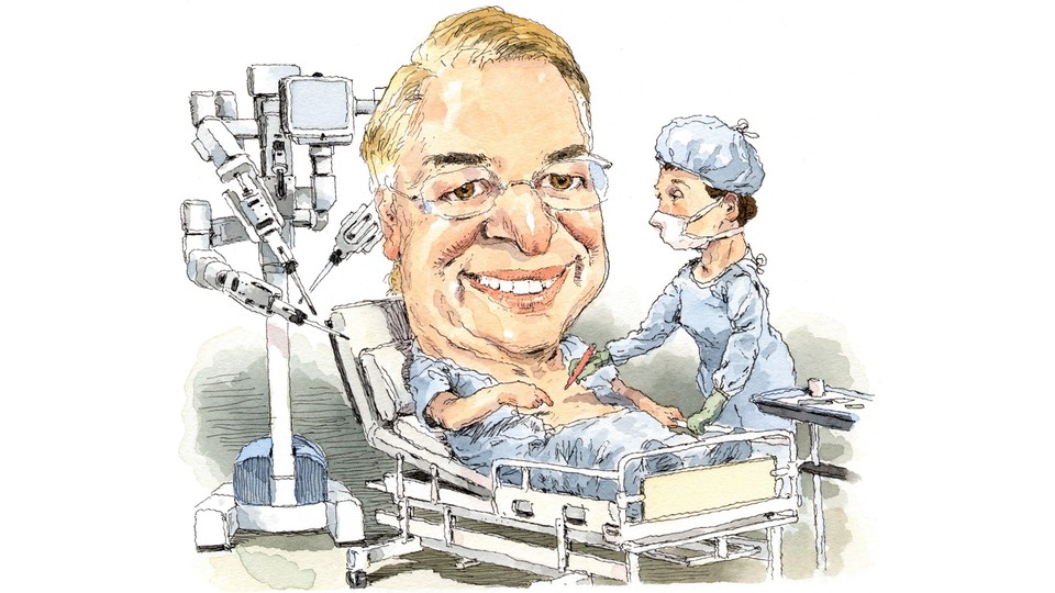
Like everyone who sees the virtual-reality room, Ramamoorthy was at first amazed. Then she was struck by how useful the images were. Inside our bellies, the intestines are a jumble of coiled tissue, resting among other organs and vital blood vessels. The twists and turns are not the same in everyone, so when a surgeon peers in, she encounters a layout that can differ from one person to the next and that, considering the coils are folded into such a small space, can be hard to sort out. In the operating room, the patient is placed on a reclining board with his head down, so that gravity eases compaction and makes the work a little easier. The first step in the procedure, under normal circumstances, is to insert a scope into the belly for a look around.
“We see kind of the lay of the land, and get a sense of what we have to do,” Ramamoorthy explained. She performs surgery with a state-of-the-art robot called the da Vinci Xi, a four-armed device that nearly fills the operating room. At the end of each arm is a narrow tube that can be inserted into the patient’s body; each insertion point is called a port. A small camera or the robot’s delicate fingers can be threaded through the tubes at these ports. Placement of the ports is crucial, because the camera and fingers that extend from them into the patient’s viscera must be set precisely in the area where Ramamoorthy intends to cut.
When using the da Vinci, Ramamoorthy does not peer directly into the patient’s body, but instead views it through a scope at the da Vinci’s workstation, which has a screen to project what the robot’s cameras see, and hand controls with which she can manipulate the robot’s fingers. The first step in Larry’s procedure would be to determine exactly where to place the ports in his belly.
Inside Transparent Larry, however, Ramamoorthy got a jump on the surgery a week early. She could see which portion of the colon would have to be removed, where it was located, and how it was shaped. All the peculiar twists and folds of Larry’s organs were displayed. She could see, near the upper-left end of his colon, where it was attached to his spleen, and where, in another spot, one of its folds pressed against his bladder—both areas of surgical risk. “You’re the doctor, not me,” Larry told her, pointing to a precise spot, “but I would start cutting here.” Pointing to another spot, he added, “And cutting here makes a good deal of sense.”
“That’s about right,” she said.
She would later calculate that this virtual inspection “probably saved us about an hour’s worth of time during surgery.” That’s a valuable advantage, she explained, because the longer a patient is under anesthesia, the more likely he is to suffer postoperative complications.
When it came time for Ramamoorthy to review the necessary consent forms with Larry, both understood the surgical plan exactly, where the danger points were, and when decisions might have to be made in the moment—not in a generic sense, as with most surgeries, but with great specificity. Larry was functioning, in a concrete sense, as his body’s CEO.
“I was really the learner and he was the teacher,” Ramamoorthy said.
Before the surgery, Larry arranged for Intuitive Surgical, which makes the da Vinci, to work with Jürgen Schulze, his colleague, to feed his 3-D images directly into the robot. This would enable Ramamoorthy to see 3-D virtual images in her scope alongside the real images of Larry’s colon from the da Vinci’s stereo high-definition camera. Days before the surgery, she told Schulze that she wanted him to participate in the procedure. When Schulze initially demurred, citing a discomfort with blood, Larry told him: “Man up.”
The scene in Larry’s operating room on November 29, 2016, looked more like a crowded booth at an engineering convention than a surgical theater. Larry’s supine body, completely draped in blue paper except for his swollen belly—tinted orange with antibacterial swabbing—was surrounded by a thicket of white, plastic-clad robotic arms and industrious doctors, nurses, and technicians. The university’s media-relations office had gotten wind of the groundbreaking effort, so a video crew was present too. Schulze was there with his laptop, manipulating the virtual version of Larry’s insides. Ramamoorthy, after making the initial incisions and inserting the rods containing the robot’s camera and fingers, sat in a corner at the controls, orchestrating the procedure. “I love it!” she exclaimed as they began. “This team is on!”
Explaining her moves as she worked, the surgeon called out from time to time for Schulze to tinker with the virtual image. “Maybe give me a lateral view of the bladder from the right side,” she said at one point.
The virtual images were so helpful, she said later, that she wishes she could have them every time she operates: “It was wonderful. It was like the difference between driving around before and after Google Maps.”
The only overtly bloody moment came when the portion of Larry’s colon was removed. It was hugely swollen, a mass of inflamed tissue the size of a melon.
About four months later, presenting his case in a lecture to the medical staff of UC San Diego Health’s Moores Cancer Center, Larry quipped, “I myself had a sort of a cameo. I played the belly in the video, sort of a Quentin Tarantino thing.”
Larry proudly passed around a plastic model of his new, streamlined colon. His symptoms had abated, and he had gone back to walking 10,000 steps a day only two weeks after his five-hour surgery. And although his blood and stool biomarkers are now back in the normal range—one of them 2,000 times lower than it was at his sickest—Larry is still on the case, trying radical shifts in his diet and tracking the effects on his microbiome. He will be 70 this year, and he hopes to find a more permanent solution to his affliction. He is still frustrated, he told me, echoing the lament of scientists everywhere, “by what I don’t know.”
Recommended Reading


Turning a two-dimensional MRI into three dimensions is not that hard, Larry told the audience at his lecture. The remaining challenge is to get more doctors to be like Ramamoorthy, and to get more engineers working in concert with them. Larry wants to build a hub on the UCSD campus to meld the isolated disciplines into a functioning whole. “We have the top-line medical people and facilities, and we have the researchers,” he said. “It’s just the social organization of getting out of the stovepipes long enough to put these kind of teams together.”
When I first met Larry Smarr, he was trying to chart a new future for the diagnosis of illnesses. Today, he’s also charting a future for the surgeries used to treat them. And he’s demonstrating—quite dramatically—what it’s like when the patient, not the doctor, is in charge.
This article appears in the March 2018 print edition with the headline “The World’s Most Body-Conscious Man.”