UK Surgeons Use Advanced 3D Camera Technology to Assist with Heart Bypass Surgery

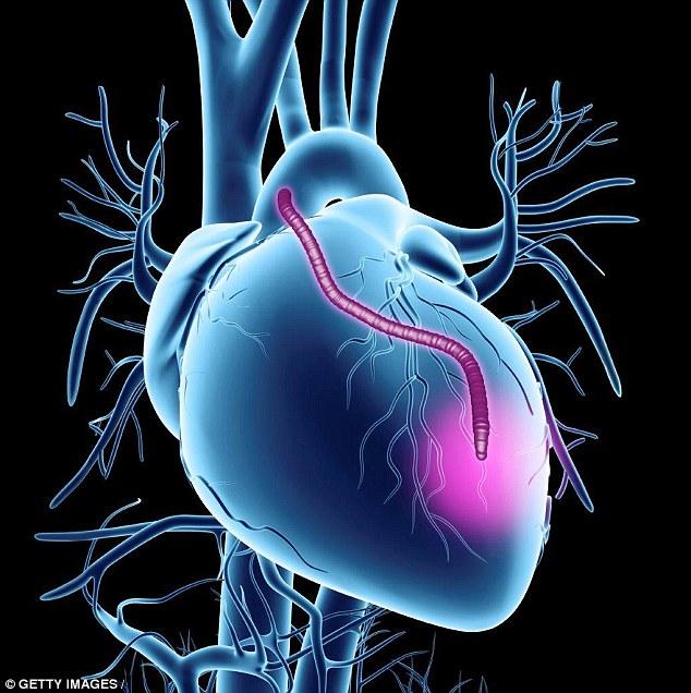
A stent being placed inside of a heart.
Coronary artery disease is a condition that develops when cholesterol and calcium deposits begin to build up inside the arteries, causing them to become narrower and restrict blood flow to the heart. The resulting chest pain from the reduced blood flow is called an angina, and is often seen as a precursor to a heart attack if the artery becomes completely blocked. One of the more common procedures used to repair these blocked arteries involves placing small metal tubes called stents inside the artery to keep it open and prevent further blockage. These procedures can typically be done relatively easily using minimally invasive keyhole, or laparoscopic, surgeries. Unfortunately stents are not always capable of repairing the blockage and a more complex, and invasive, surgery needs to be performed.
If the block is too extreme, then doctors will take a blood vessel from behind the breastbone, or occasionally from a leg or an arm, and use it to bypass the blockage in the heart. The results are usually completely effective, however the surgery is quite invasive and requires the breastbone to be cut open in order to expose the heart. That kind of incision will often take up to six months to fully heal, and many times the patient already isn’t in the best of health, making the surgery quite risky. Doctors have tried using laparoscopic surgeries, however the 2D image that are sent back from the camera often makes it difficult to judge where incisions need to be made. Using a laparoscopic procedure turns the four-hour surgery into a six-hour surgery, and generally can’t be used for more difficult surgeries.
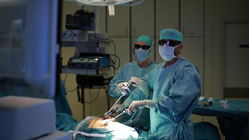
Using a thoracoscope to operate internally.
However with the rapid advancement of 3D camera technology, doctors at King’s College Hospital in London, UK have begun exploring using 3D imaging to perform more complex surgeries laparoscopically. Earlier in the year 62-year-old Bruce Fancourt from Kent was told that one of his arteries had become 96% blocked and the only way to repair it was with a bypass operation. But rather than cut open his chest, or risk a 2D laparoscopic procedure, a team of NHS hospital cardiothoracic surgeons led by Dr. Ranjit Deshpande used new 3D cameras in place of the old 2D system and successfully repaired the artery. The 3D system was not only less invasive and safer, but it dramatically reduced the amount of time that was required to perform the surgery down to two hours.
“3D technology means we still perform keyhole surgery, with all the same benefits for the patient, but in such a way that we can visualise the patient’s anatomy as if we were doing open heart surgery. This is a major step forward, and I am confident more patients like Mr Fancourt will benefit in the future,” said Dr. Deshpande.

The 2 cameras used to create the 3D images.
The 3D technology works a lot like the 3D projection systems in movie theaters, and requires surgeons to wear a special pair of glasses in order to see the images properly. Along with the surgical tools used to perform the surgery, a tool with two cameras, each at right angles to each other, called a thoracoscope is inserted into a small incision in the patient’s chest. The images from the two cameras are transmitted to a screen in the operating room where they are superimposed on top of each other. The 3D glasses allow the surgeons to experience the inside of the patient’s anatomy in full three dimensions, as if they had actually opened the patient’s chest up. Using the thoracoscope, the surgical team can see the anatomy from all angles and perform the procedure faster and more accurately.
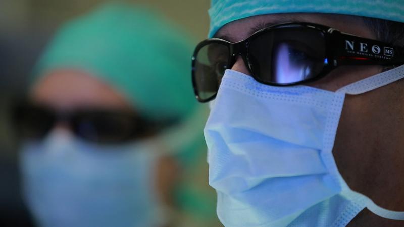
Special glasses are used to view the patient’s anatomy in 3D.
The process is called an Endoscopy Assisted Coronary Artery Bypass Surgery, and it is being used on patients who need one or two bypass grafts. However as the NHS surgeons become more experienced they expect to use it on triple or quadruple bypass grafts as well. The NHS also hopes to use the 3D technology during more complex heart surgeries like heart valve repairs and repairing congenital heart problems like holes in the heart. If the surgery is performed correctly, the grafts can stay in place for up to 20 years without any problems, and according to the Daily Mail, the entire procedure only costs the NHS £9,000 ($1320).
While the 3D thoracoscope technology had been used for other procedures at King’s College Hospital, it was only for relatively minor surgeries like gall bladder and prostate operations. The surgery on Mr. Fancourt was the first time that the technology was ever used in the UK for a heart bypass. Because the surgery was significantly faster and less invasive, Mr. Fancourt was out of the hospital in just four days, and had almost fully recovered within two weeks. Discuss this amazing technology further over in the 3D Imaging for Heart Surgery forum over at 3DPB.com.
Subscribe to Our Email Newsletter
Stay up-to-date on all the latest news from the 3D printing industry and receive information and offers from third party vendors.
You May Also Like
Further Understanding of 3D Printing Design at ADDITIV Design World
ADDITIV is back once again! This time, the virtual platform for additive manufacturing will be holding the first-ever edition of ADDITIV Design World on May 23rd from 9:00 AM –...
3D Printer Maker EVO-tech Reborn as NEVO3D — Once More With Feeling
EVO-tech was a 3D printing service and original equipment manufacturer established in 2013 and based in Schörfling am Attersee, Austria. The company produced high-quality material extrusion systems featuring linear bearings,...
3D Systems Brings 3D Printed PEEK Cranial Implant to the U.S. with FDA Clearance
For more than 10 years, 3D Systems (NYSE:DDD) has worked hand-in-hand with surgeons to plan over 150,000 patient-specific cases, and develop more than two million instruments and implants from its...
CDFAM Returns to Berlin for Second Annual Symposium
The second CDFAM Computational Design Symposium is scheduled for May 7-8, 2024, in Berlin, and will convene leading experts in computational design across all scales. Building upon the first event...