Article

TLR-2 Signaling Promotes IL-17A Production in CD4+CD25+Foxp3+ Regulatory Cells during Oropharyngeal Candidiasis

Department of Biological Sciences, School of Dental Medicine, Case Western Reserve University, Cleveland, OH 44106, USA
*
Author to whom correspondence should be addressed.
Pathogens 2015, 4(1), 90-110; https://doi.org/10.3390/pathogens4010090
Submission received: 4 February 2015 / Revised: 5 March 2015 / Accepted: 11 March 2015 / Published: 17 March 2015
(This article belongs to the Special Issue Candida Albicans Infections)

Abstract

:
Recent studies show that CD4+CD25+Foxp3+ regulatory cells (Tregs) produce effector cytokines under inflammatory conditions. However, the direct role of microbial agents that serve as toll-like receptor (TLR) ligands in the induction of effector cytokines in Tregs is less clear. Here we show that CD4+Foxp3+Tregs produce the effector cytokine IL-17A during oropharyngeal candidiasis (OPC) and inflammatory bowel disease in a TLR-2/Myd88 signaling dependent manner. TLR-2 ligands promote proliferation in Tregs in the presence and absence of TCR signals and inflammatory cytokines in vitro. The proliferation is directly dependent on TLR-2 expression in Tregs. Consistent with this, Tlr2−/− mice harbor fewer thymically derived Tregs and peripheral Tregs under homeostatic conditions in vivo. However, under Th17 inducing conditions, IL-6 and TLR-2 signaling both in Tregs as well as antigen presenting cells (APC) are critical for maximal ROR-γt and IL-17A up-regulation in Foxp3+ Tregs. The minimal and transient loss of Foxp3 expression and suppressive properties are due to the presence of IL-6 in the milieu, but not the direct effect of TLR-2 signaling in Tregs. Taken together, our data reveal that TLR-2 signaling promotes not only proliferation, but also IL-17A in Tregs, depending on the cytokine milieu. These IL-17A producing Tregs may be relevant in mucosal infections and inflammation.

Graphical Abstract

1. Introduction

Tregs play important roles in dominant tolerance and immune homeostasis [1,2], and are targeted for human immunotherapy. Therefore, it is essential to study how Tregs precisely respond under steady state conditions and inflammatory conditions. Although Tregs are conventionally non-producers of inflammatory cytokines, recent reports document the presence of interleukin (IL)-17+Foxp3+ cells in mucosal environments in mice as well as in several human diseases [3,4,5,6,7]. This novel subset of cells likely represents an intermediate differentiation stage between Th17 cells and Tregs. However, the mechanism by which they are induced, and their exact functions remain unknown [6,8]. TLR-2 recognizes bacterial lipopeptides, lipoteichoic acid from Gram-positive bacteria, zymosan from yeast cell walls, and the synthetic lipoprotein Pam3CSK4 [9]. Toll like receptors (TLR)s are generally expressed by the innate immune cells and APC, and play critical roles in host defense [10,11]. T lymphocytes, including Treg cells demonstrate Toll-like receptor signaling as well [12]. TLR-2 polymorphisms are associated with changes in neonatal Treg numbers as well as allergies and atopic dermatitis [13]. In vitro, Pam3CSK4 directly acts on CD4 cells and enhance their IL-17 production during Th17 differentiation [14]. However, research on TLR-2 signaling in Tregs has produced some varied results. Some studies show that TLR-2 ligands reduce Foxp3 expression in Tregs, and reverse their suppressive functions [15,16]. Others show that TLR-2 ligands improve the survival of Tregs without reversing their suppressive functions [17]. By inducing peripheral Tregs (pTregs), TLR-2 signaling also promotes gut tolerance and not inflammation [18]. Though some of the above studies imply that suppression by Tregs may be overcome by TLR ligand induced DC maturation, and pro-inflammatory cytokine induction in effector cells [19,20], whether some of the effects were directly mediated by TLR-2 signaling in Tregs, remains incompletely defined. Additionally, some data suggest that Tregs may play a deleterious role during acute disseminated candidiasis [21], however we as well as others have previously shown their protective functions during acute OPC and reinfection [22,23,24]. Whether Tregs themselves produce effector cytokines was not investigated at the time. In this study, we have shown that during OPC and under Th17 inducing conditions in vitro, TLR-2 ligands promoted proliferation and IL-17A induction in Foxp3+Tregs. Increased proliferation was dependent on TLR-2 expression in Tregs. However, IL-17A production was dependent on TLR-2 signaling in Tregs and APC, as well as IL-6 in the milieu. Under Th17 inducing conditions, though the presence of IL-6 in the milieu minimally reduced Foxp3 expression, TLR-2 ligand stimulation did not directly reduce Foxp3 expression. The TLR-2 activated Tregs, which included IL-17 producing Tregs, retained suppressive activity. Taken together, we have identified the direct role of TLR-2 ligands in promoting proliferation and IL-17A production in Tregs, without affecting their suppressive functions in vitro and in vivo.

2. Results

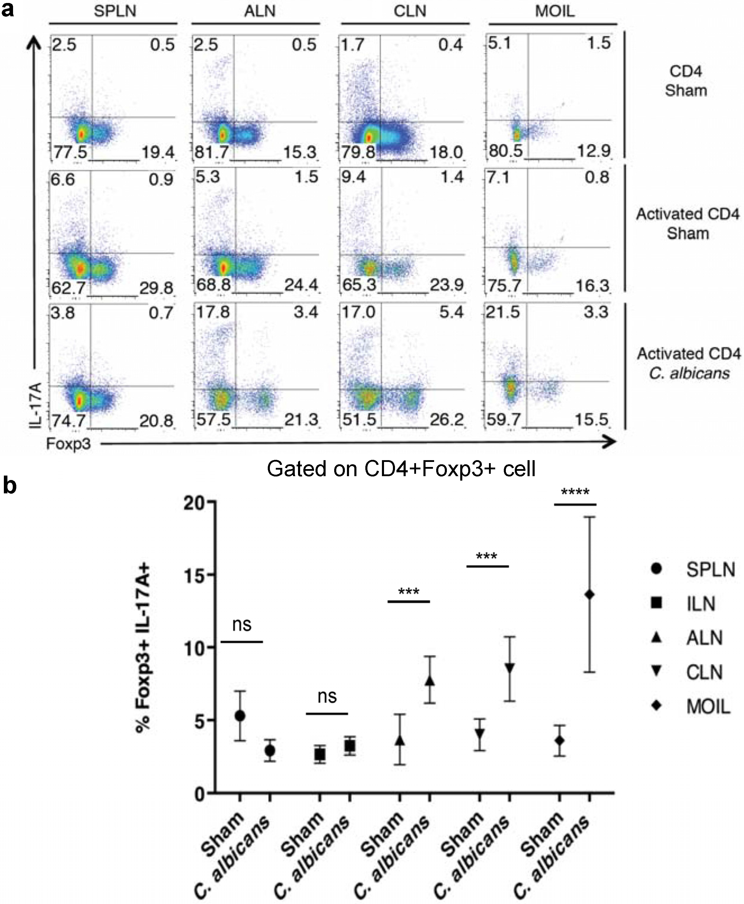
2.1. CD4+CD25+Foxp3+ Tregs Express IL-17A During Oral C. Albicans Infection and Inflammatory Bowel Disease (IBD) in Vivo

We and others have previously shown that IL-17A is critical for anti-Candidal host resistance [22,25], and Tregs enhance acute effector Th17 responses and fungal clearance during OPC [22,23]. Therefore we monitored the expression of IL-17A in Foxp3+ Tregs in the same OPC infection model by orally infecting the Foxp3GFP reporter mice with C. albicans. On day 2 after infection, we isolated the cells from spleen, lymphnodes and mouse oral lamina propria and intraepithelial cells (MOIL) isolated from tongue and palatal tissues [26]. Both CD4+Foxp3 effector cells and CD4+Foxp3+ Tregs responded to infection by expressing IL-17A in the draining axillary lymph nodes (ALN), cervical lymph nodes (CLN) and MOIL in infected mice (Figure 1a). Although there were a few Foxp3+IL-17A+ cells in sham infected mice, CD44high (activated) CD4 cells showed a substantial increase in Foxp3+IL-17A+ cells only in infected mice. However, there was no increase of these cells in spleen (Figure 1a). We further confirmed the induction of Foxp3+IL-17A+ cells by gating on CD4+Foxp3+ cells only (Figure 1b,c). Although a fraction of Tregs produced IL-17A on d1 and d2 after infection as shown previously [22], Tregs still increased IL-17A in effector cells, decreased fungal burden, and modulated immunopathology and weight loss in Treg recipients during OPC on d5 (data not shown, [22]).
Figure 1. CD4+Foxp3+ Tregs express IL-17A during C. albicans infection in vivo. (a) Foxp3GFP reporter mice were infected with PBS sham or C. albicans. On day 2 after infection, the cells from spleen, lymph nodes, and MOIL were isolated and restimulated for cytokine analyses. Flow cytometric plots showing Foxp3 and IL-17A expression, gated on CD4+ cells (top), and CD4+CD44high activated CD4 cells (middle and bottom rows). (b) Cells were isolated as in “a”. Flow cytometric analyses of IL-17A expression, gating on CD4+Foxp3+ cells in sham or C. albicans infected mice. Data represent triplicate experiments.
Pathogens 04 00090 g001
Human mucosal Foxp3+Tregs express IL-17A in inflammatory conditions, such as periodontitis, psoriasis and IBD [4]. Therefore, we next tested whether IL-17A+Foxp3+ cells are induced during IBD in mice. We adoptively transferred CD45.1 Th17 cells into CD45.1 Rag1−/− mice to initiate IBD [22,27]. Two weeks after IBD induction, we injected CD4+CD25+GFP+ Tregs, derived from congenic CD45.2 Foxp3GFP reporter mice. Five days after Treg injection, we isolated spleen, mesenteric lymph nodes (MLN) and mouse gut intraepithelial and lamina propria leukocytes (MGIL) and measured the IL-17A induction upon restimulation in vitro. We gated on CD4+Foxp3CD45.1+effector cells and CD4+Foxp3+CD45.1-Tregs to examine IL-17A induction in these cells. We observed that Tregs were able to produce IL-17A, especially in MLN and LP during inflammation in vivo (Figure S1a). Despite IL-17A production in Tregs, the injected Tregs still modulated IBD and weight loss in Treg recipients (Figure S1b). Taken together, these data demonstrate that infections and inflammatory conditions can induce IL-17A production in a fraction of Foxp3+Tregs in vivo, but do not affect their immunomodulatory functions.

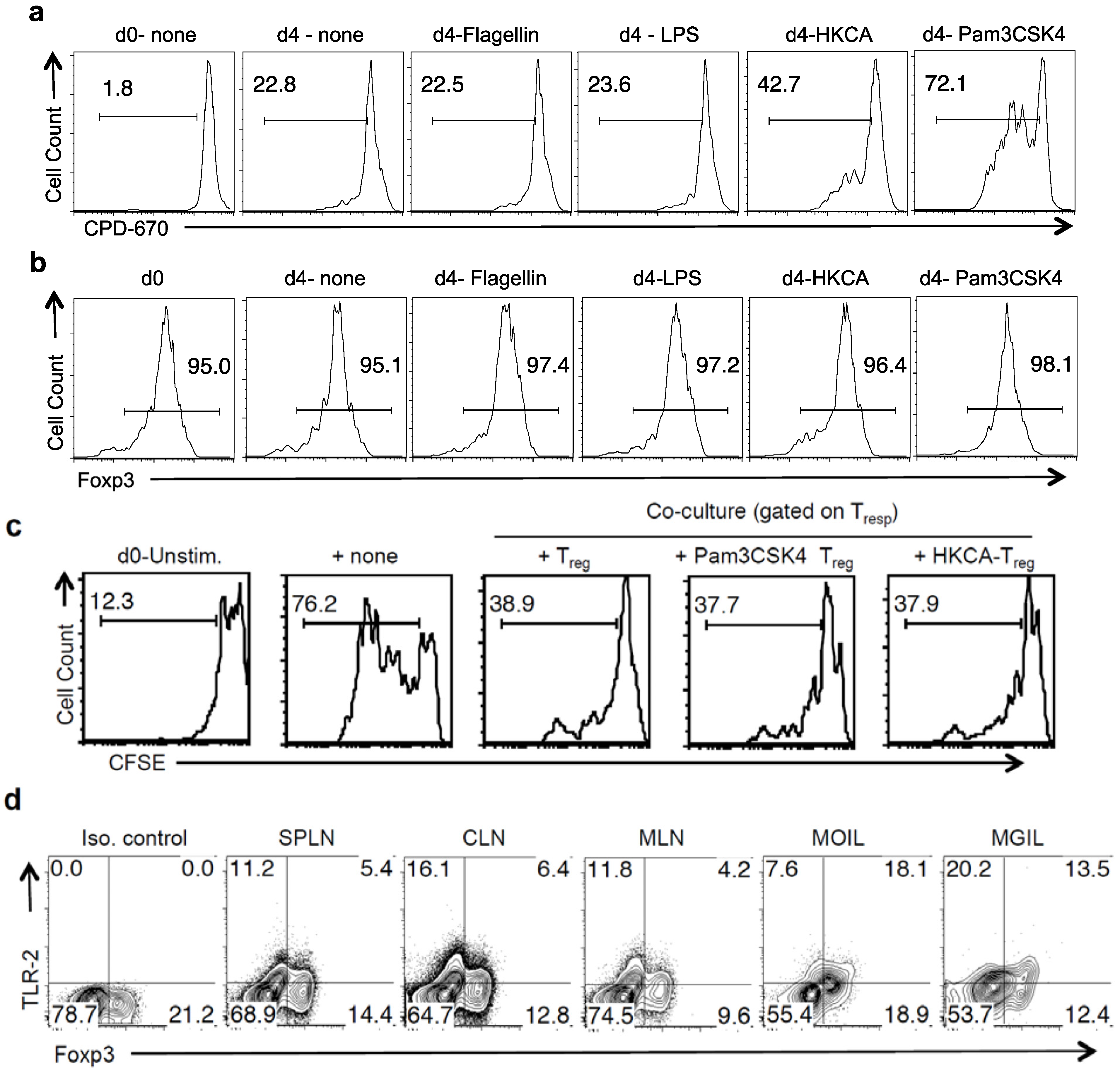
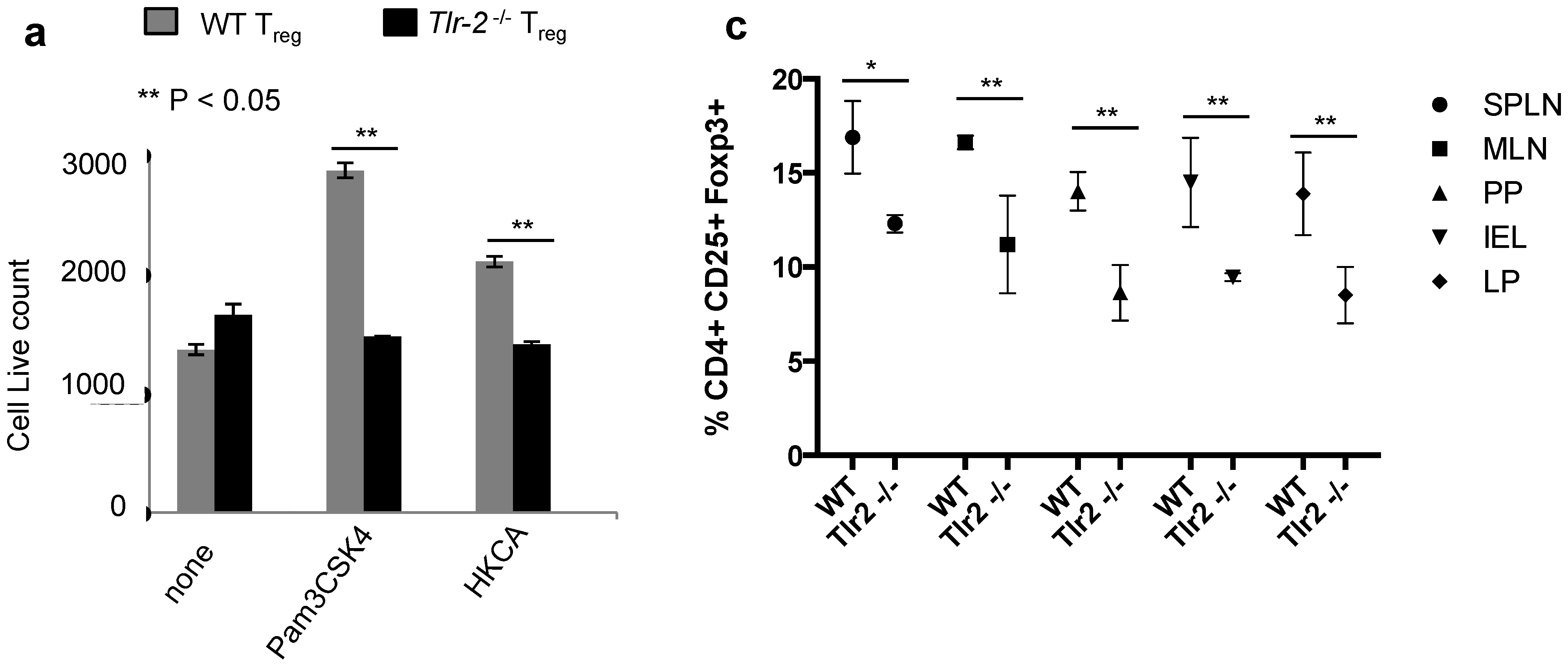
2.2. TLR-2 Ligands Directly Induce Proliferation of Tregs Independently of TCR Activation

As C. albicans activates TLR-2 and dectin signaling that promote Th17 responses [28], we examined whether TLR-2 ligands and dectin ligands induced IL-17A in Tregs in vitro. We sought to investigate the effects of TLR-2 ligands on highly purified CD4+CD25+Foxp3+ Tregs under non-inflammatory and inflammatory conditions. First we examined whether TLR ligands impacted Tregs in the absence of TCR activation, but with IL-2 alone. IL-2 stimulation of Tregs in the absence of TCR ligation would be akin to non-inflammatory conditions and would induce homeostatic proliferation in Tregs [29,30,31]. We isolated CD4+ CD25+Foxp3GFP+ cells (Treg) and control CD4+CD25Foxp3GFP−CD44l°w naïve T cells (Tcon) from Foxp3GFP reporter mice (Figure S2), and stimulated them with IL-2 [30]. This condition also prevented the proliferation of possible contaminating Tcons among Tregs, which expand vigorously with TCR stimulation in cultures. We labeled Tregs with cell proliferation dye-670 (CPD-670) and tested their viability and proliferation with and without various TLR ligands. As expected, naïve Tcon cells died under these conditions in the absence of TCR activation (data not shown). However, IL-2 stimulation alone maintained Treg viability by virtue of constitutive IL-2 receptor expression on Tregs. On day 4 after stimulation, heat killed Candida albicans (HKCA), a natural Tlr-2/Dectin ligand, and Pam3CSK4, a TLR2/6 ligand increased the proliferation of Tregs (Figure 2a). Other TLR ligands, such as Poly A:U (TLR-3) (data not shown), LPS(TLR-4) and Flagellin (TLR-5), showed no effect (Figure 2a). As TLR-2 ligands have been shown to transiently reduce Foxp3 mRNA expression of TCR activated Tregs [15], we measured Foxp3 expression of Tregs. HKCA and Pam3CSK4 did not reduce Foxp3 expression in Tregs (Figure 2b). To examine their suppressive capacity, we washed the Tregs to remove the TLR-2 ligands, and stimulated them with α-CD3 and APC along with freshly isolated, carboxyfluorescein di-acetate succinimidyl ester (CFSE) labeled CD4+CD25 responding cells (Tresp) in co-cultures. Tresps that were stimulated alone without Tregs showed increased proliferation (76.2%) compared to Tresps that were stimulated with Tregs (38.9%) on day 4 after stimulation (Figure 2c). Tregs that were previously stimulated with IL-2 alone, or with Pam3CSK4 or HKCA, were capable of suppressing the proliferation of Tresps (Figure 2c). These data show that TLR-2 ligands, along with IL-2 can induce proliferation of Tregs in the absence of TCR signals, without affecting their suppressive capacities. Because Tregs responded to TLR-2 ligands without TCR activation, we hypothesized that they might express TLR-2 protein ex vivo. We found that both Foxp3 and Foxp3+ CD4+ cells from spleen and lymph nodes expressed TLR-2 ex vivo (Figure 2d). Interestingly, TLR-2 expression was markedly increased in mucosal Tregs found among MOIL and mouse gut intraepithelial and lamina propria cells (MGIL), implying an important role of TLR-2 in mucosal Tregs.
Figure 2. Tregs proliferate in response to TLR ligands and IL-2 independently of TCR. (a) CPD-670 dilution (proliferation) of Treg cells, with indicated TLR ligands added at the beginning of stimulation; (b) Histograms of Foxp3 expression of live Tregs that were stimulated as in “a”; (c) Tregs stimulated with or without TLR-2 ligands as in “a”, were isolated and washed after two days. They were restimulated with fresh CFSE-labeled CD4 naive responder (Tresp) cells in co-cultures. Some Tresp cells were not stimulated (d0-unstim) or stimulated alone (+ none). Tresp proliferation is shown; (d) Foxp3 and TLR-2 expression in CD4 T cells isolated ex vivo from indicated tissues. (ad) represent three independent experiments.
Pathogens 04 00090 g002

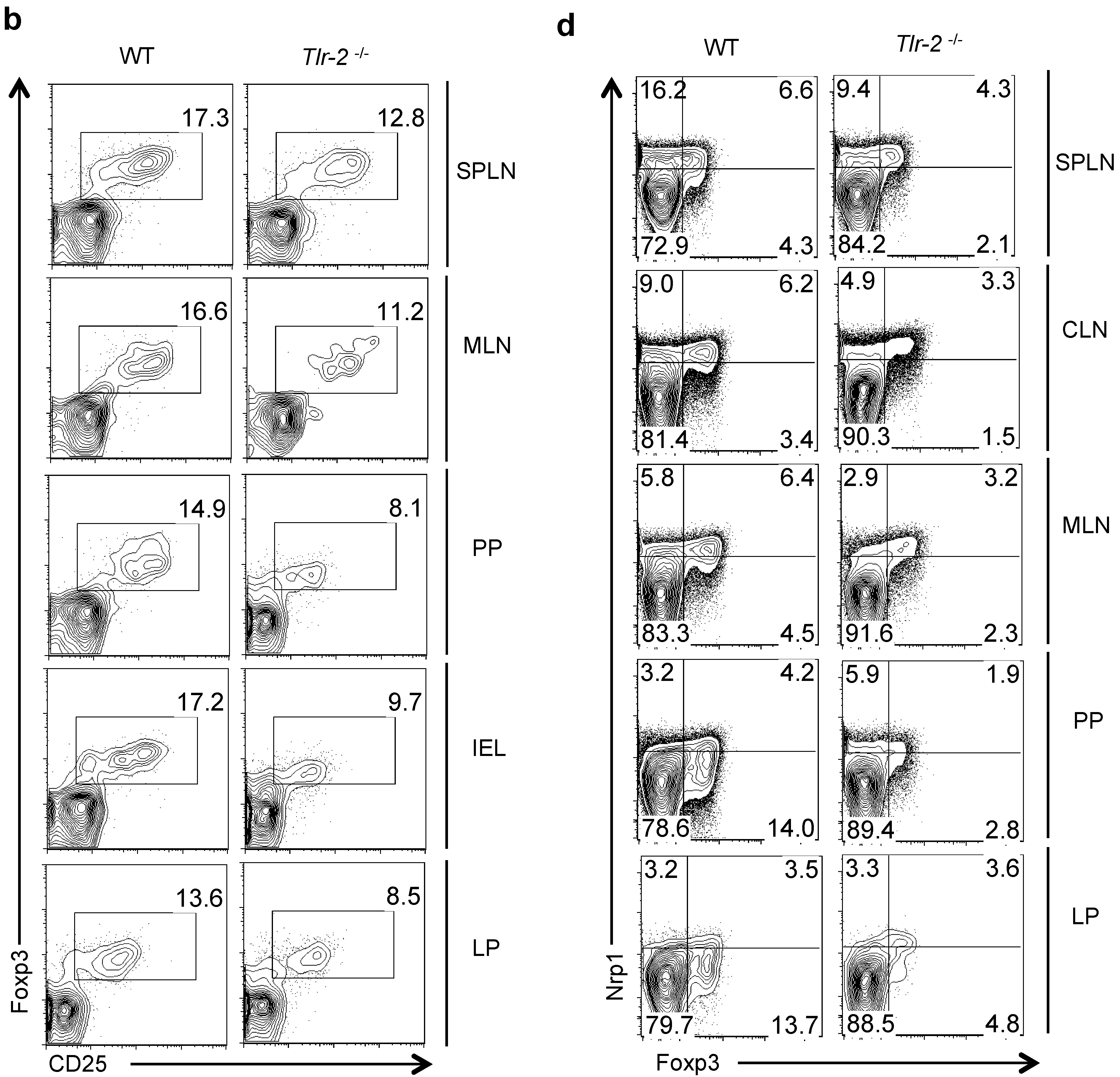
2.3. TLR-2 Ligand Mediated Proliferation in Tregs is Directly Dependent on TLR-2 Expression on Tregs

We then sought to confirm that proliferation induced by TLR-2 ligands was dependent on TLR-2 expression in Tregs. We isolated Tregs from the spleens of WT or Tlr-2−/− mice and stimulated with IL-2 and TLR-2 ligands. On day 3 after stimulation, HKCA and Pam3CSK4 increased the Treg cell numbers in WT Tregs but not in Tlr-2−/− Tregs, showing that they induced proliferation in TLR-2 dependent manner in Tregs (Figure 3a). If TLR-2 signaling induced proliferation of Tregs in the absence of TCR ligation or inflammatory cytokines, TLR-2 agonists in gut commensal microbes and other endogenous ligands may also promote Treg proliferation under homeostatic conditions in vivo. To test this idea, we examined the frequency of CD4+CD25+Foxp3+ Tregs in various organs, including SPLN, MLN, payer’s patches (PP), gut lamina propria (LP), and the gut intra epithelial lymphocytes (IEL) in WT and Tlr-2−/− mice, using flow cytometry. We found that the frequency of CD4+CD25+Foxp3+ cells was significantly reduced in Tlr-2−/− mice when compared to the WT mice (Figure 3b,c). A previous study showed that the frequency of CD4+CD25+ cells was lower in the peripheral blood of Tlr-2−/− mice than in WT mice [21]. However, neither Foxp3 expression nor the frequency of thymically derived Tregs (tTregs) and peripheral Tregs (pTregs) was examined. Therefore we evaluated the expression of neuropilin-1 (Nrp-1), a marker on tTregs in peripheral organs and mucosal tissues. The frequency of both the Treg populations were decreased slightly in SPLN, but dramatically in CLN, MLN, PP and LP of Tlr-2−/− mice, when compared to the WT mice (Figure 3d). Interestingly, in payer’s patches and the intestinal lamina propria, the reduction of Tregs in Tlr-2−/− mice was more pronounced in the Nrp-1 pTreg compartment (Figure 3d). These results highlight the importance of TLR-2 ligands in promoting tTreg and pTreg homeostatic proliferation under steady-state conditions in vivo.
Figure 3. Pam3CSK4 and HKCA induce proliferation of Tregs in a TLR-2 dependent manner. (a) Live cell counts (PIneg and FSChigh) of Treg cells from WT (grey) or Tlr-2−/− (black) mice stimulated with various TLR-2 ligands for three days as in “2a”. CD4+CD25+Foxp3+ (b), or CD4+ Foxp3+Nrp1+ (d), frequencies in WT (left) and Tlr-2−/− (right) cells isolated ex vivo. (c) Statistical analyses showing Treg frequencies in WT and Tlr-2−/− mice, as determined in “b”.
Pathogens 04 00090 g003aPathogens 04 00090 g003b

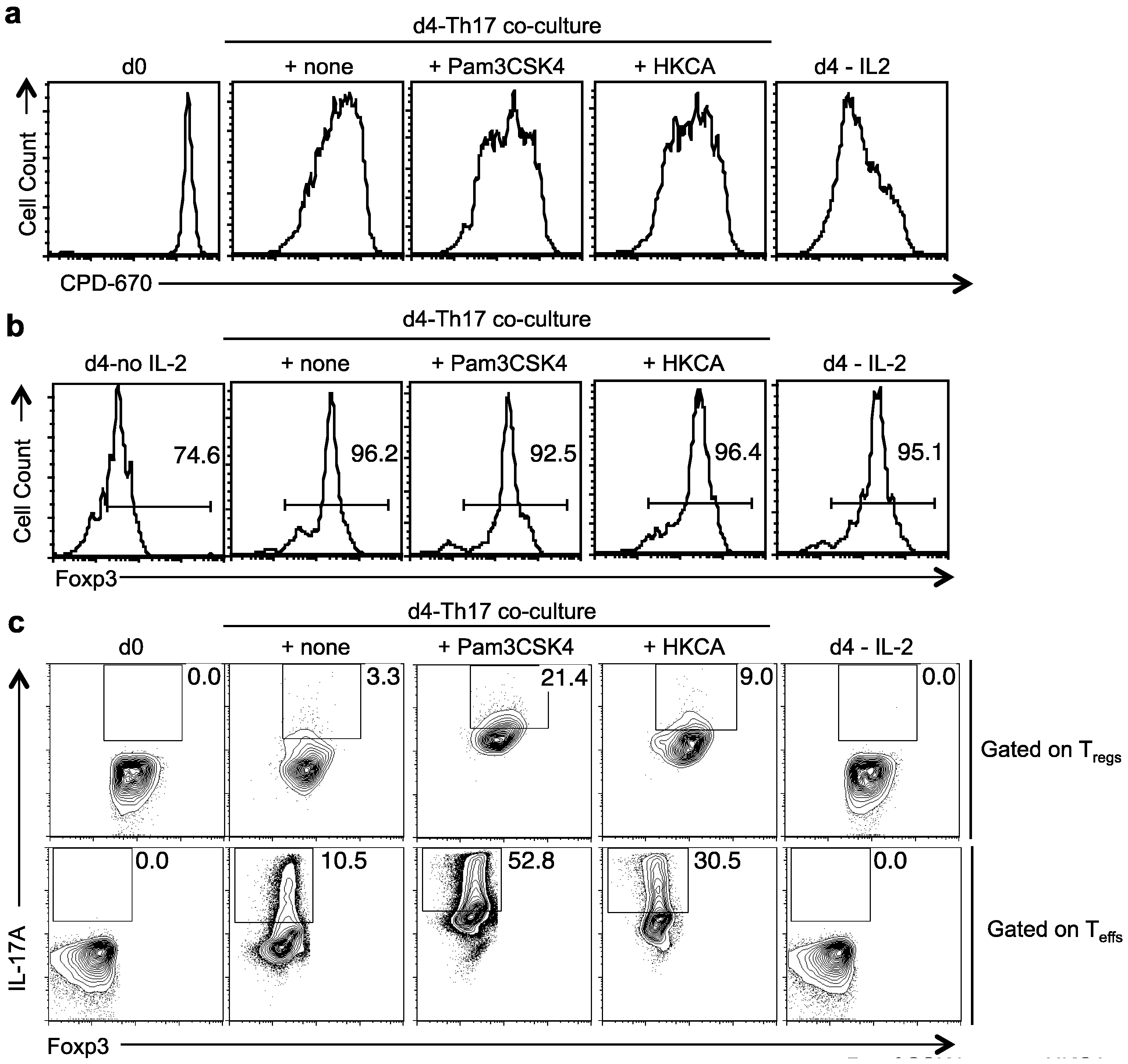
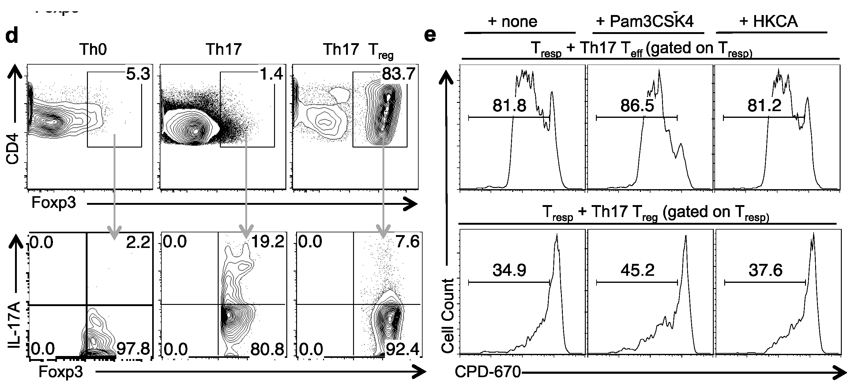
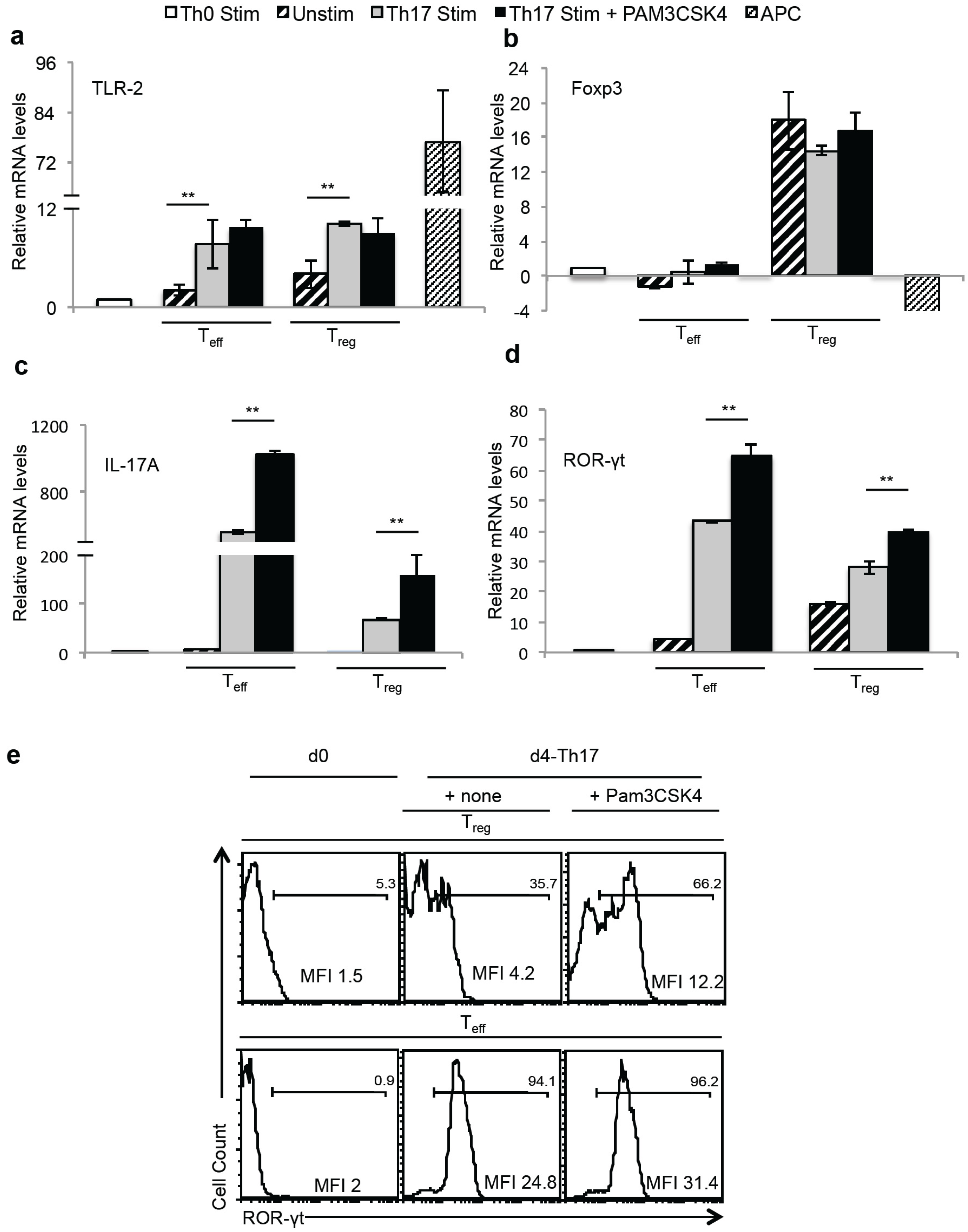
2.4. TLR-2 Ligands Did Not Reduce Foxp3 Expression, but Induced IL-17A in Foxp3+ Tregs under Th17 Conditions in Vitro

We then sought to investigate whether TLR-2 ligands may promote IL-17A in Tregs stimulated under Th17 inducing conditions in vitro. Although TLR-2 ligands have been shown to reduce Foxp3 expression and suppressive properties in Tregs [15], whether IL-17A is induced in Tregs is unknown. However, Strober and colleagues have shown that Tregs produce IL-17A in Th17 inducing conditions in vitro [32]. Therefore, we stimulated Tregs in co-cultures along with Tcons at a ratio of 1:10 under Th17 inducing conditions. Tcon cells proliferated in to Th17 effectors (Teff). As controls, we stimulated Tregs in Th0 conditions. To distinguish Tregs and Teff in co-cultures, we labeled Tregs using CPD670 in addition to CD45.2 congenic marker on Tregs in co-cultures (Figure S3a). We gated on CPD670+CD45.2+Tregs (Figure S3a), and examined their proliferation, Foxp3 expression and IL-17A expression. Four days after stimulation, Pam3CSK4 and HKCA increased the proliferation of Tregs (Figure 4a). Although ~20% of the cells lost Foxp3 expression completely, many of these cells were non-proliferating Tregs, which did not dilute CPD670 (Figure S3b). These cells were found in the presence and the absence of TLR-2 ligands (Figure S3b), likely due to the fact that IL-6 that can transiently reprogram Tregs to lose Foxp3 expression [22,33]. However, when proliferating Tregs were examined, TLR-2 ligands did not lead to reduction in Foxp3 expression (Figure 4b). As expected, in the absence of IL-2, Tregs showed poor survivability and reduced Foxp3 expression ([30], Figure 4b). We also observed that a small fraction of the Foxp3+ cells (~3%) consistently expressed IL-17A (Figure 4c). However, TLR-2 ligands increased the frequency and the levels of IL-17A substantially (Figure 4c, upper panel). We did not observe IL-17A expression in control Th0 stimulated Tregs (Figure 4c, upper panel). To verify the specificity of the staining, we used Tregs from Il17a-/-cre mice, which barely showed IL-17A expression (Figure S4). Under Th0 and Th17 polarizing conditions, a small fraction of Teffs also induced Foxp3 expression (iTregs) (Figure 4d, upper panel). iTregs that were induced in Th0 conditions did not express IL-17A, while iTregs induced under Th17 polarizing conditions up-regulated IL-17A, similar to natural Tregs (Figure 4d). These data show that both in vitro induced iTregs and natural Tregs isolated from mice are capable of producing IL-17A under Th17 inducing conditions.
Figure 4. TLR-2 ligands induce proliferation but not plasticity in Tregs stimulated under Th17 conditions. CPD-670 dilution (proliferation) (a), or Foxp3 expression (b), of Treg cells co-cultured with conventional CD4+ T cells under Th17 conditions for four days, with or without Pam3CSK4, HKCA, or by themselves without (d4-no IL-2) or with IL-2 (d4-IL-2). CD45.2 Tregs are gated in the analyses. (c) Tregs were stimulated as in “a”. Foxp3 and IL-17A expression of cells gated on CPD670+CD45.2 Tregs (upper panel), or CPD670-Foxp3-CD45.1 Teff (lower panel) in co-cultures. (d) Foxp3 and CD4 expression (upper panel), of naïve cells that were stimulated under Th0 (Th0) or Th17 (Th17) conditions, or Tregs that were stimulated under Th17 (Th17 Treg) conditions. Foxp3 and IL-17A expression (lower panel), of the Foxp3+ cells in the upper panel. (e) Proliferation suppression of the CD4 responder cells (as in “2c”) co-cultured with GFP+ Treg cells isolated from cultures stimulated as in “a”. Data represent three independent experiments.
Pathogens 04 00090 g004aPathogens 04 00090 g004b
As we have shown previously, Tregs did not suppress effector Th17 cells (Teff) because of IL-6 and excess IL-2 in Th17 polarizing milieu [22], both in the presence or absence of TLR-2 ligands (data not shown). We then sought to determine whether Tregs retain their suppressive activity after the withdrawal of Th17 cytokines and TLR-2 agonists. Therefore we stimulated Tregs under Th17 inducing conditions, in the presence or absence of Pam3CSK4 and HKCA. After 4 days, we washed the Tregs (Th17 Tregs) and co-cultured them with CPD-670 labeled CD4+CD25naïve responder cells (Tresp), in the presence of α-CD3 and APC [34]. As controls, we used Teffs (Th17 Teff) that were previously stimulated under Th17 inducing conditions in the presence or absence of TLR-2 ligands, and co-cultured them with Tresps. As determined by Tresp CPD-670 dilution, we found that Th17 Tregs stimulated with TLR-2 agonists were capable inducing proliferation suppression in Tresps (Figure 4e). These results demonstrate that Tregs produced IL-17A and lost their suppressive capacity only transiently in the presence of Th17 cytokines in the milieu. However, they were capable of suppressing CD4+ T cells upon pro-inflammatory cytokine and TLR-2 ligand withdrawal.
To further validate the role of TLR-2 ligands in induction IL-17A in Foxp3+Tregs, we assessed the relative mRNA levels of Tlr-2, Foxp3, IL-17A and ROR-γt on d2 after stimulation. We used Th0 stimulated CD4+CD25 Foxp3GFP− cells as the normalizing control, unstimulated CD4+CD25 Foxp3GFP− cells, and CD90 depleted APC as additional controls. Compared to Th0 cells, both CD4 effector cells and Tregs had higher levels of Tlr-2 mRNA expression under Th17 inducing conditions (Figure 5a). As expected, Foxp3 mRNA expression was observed only in Tregs and was unchanged between un-stimulated Tregs and Th17 Tregs that were stimulated with and without Pam3CSK4 (Figure 5b). However, Th17 Tregs expressed IL-17A and ROR-γt that were further up-regulated by Pam3CSK4 (Figure 5c,d). Similar to mRNA expression, Tregs stimulated with Pam3CSK4 showed ROR-γt protein up-regulation, although at lower levels (MFI ~4–12) when compared to Teff (MFI ~24–31) (Figure 5e). Taken together, these results validate that although TLR-2 ligands induced proliferation and increased IL-17A and ROR-γt expression among Foxp3+Tregs, they do not reprogram the cells or affect the stability of Foxp3 expression.
Figure 5. Tregs up-regulate TLR-2, Foxp3, IL-17A and ROR-γt in the presence of Pam3CSK4 and Th17 conditions in vitro. Naïve cells or Tregs were stimulated under Th0 (Th0 stim) or Th17 (Th17 stim) conditions as in “4d”, with or without Pam3CSK4. Unstimulated CD4 T cells (Unstim) and CD90 depleted APC activated with Pam3CSK4 were used as controls (APC). Two days later, RNA was isolated from CD90 sorted T cells from Th17 cultures, and relative mRNA levels of Tlr-2 (a), Foxp3 (b), IL-17A (c) and ROR-γt (d) were assessed using q-PCR. (e) ROR-γt expression of the cells, gated on Foxp3+ CD45.2 Tregs (upper panel), or Foxp3-CD45.1 Teff (lower panel) in the co-cultures is shown. Geometric mean fluorescence intensities (MFI) are shown.
Pathogens 04 00090 g005

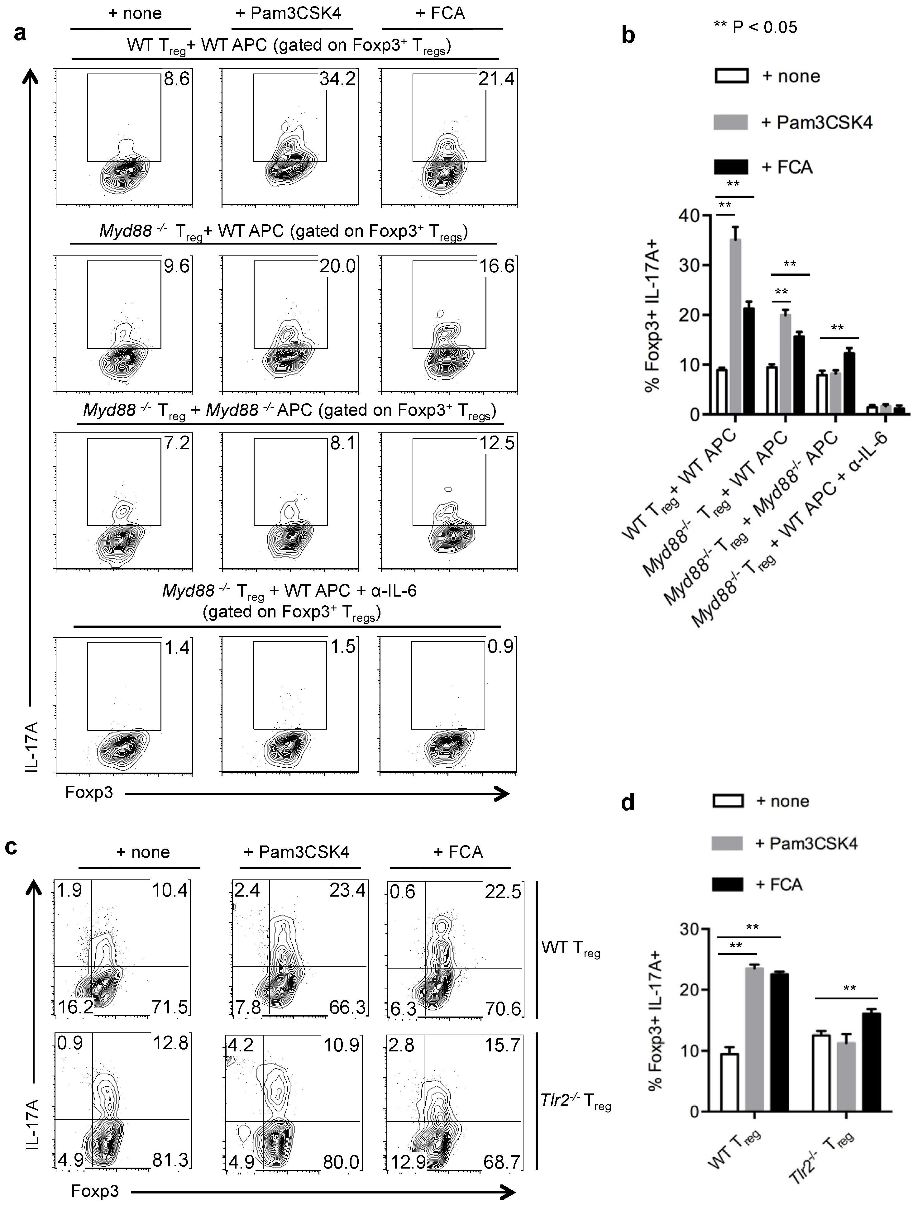
2.5. TLR-2 Ligands Induced IL-17A Production Is Only Partially Dependent on TLR-2 Expression in Tregs

We next investigated the requirement of TLR-2 receptor in induction of IL-17A in Foxp3+Tregs. Myd88 is the critical adaptor molecule downstream of TLR-2 ligand stimulation [35], and therefore we examined whether Myd88 is required for IL-17A up-regulation. We isolated WT or Myd88−/− Tregs and stimulated them under Th17 inducing conditions in the presence of WT APC with or without Pam3CSK4 and formaldehyde fixed Candida albicans hyphae (FCA). We found that the absence of Myd88 signaling in Tregs slightly reduced TLR-2 dependent IL-17A production in the presence of WT APC (Figure 6a, top two panels, 6b). We then stimulated Myd88−/− Tregs with Myd88−/− APC in the presence or absence of TLR-2 ligands. Absence of Myd88 in APC further reduced the IL-17A induction almost to the basal levels in Tregs (Figure 6a, third panel, 6b). Previous studies have shown TLR activation in APC can promote IL-6, a crucial cytokine for IL-17A induction in CD4 T cells [16,19]. Accordingly, when we stimulated Myd88−/− Tregs with WT APC in the presence of α-IL-6 neutralizing antibody, IL-17A was completely abolished in Tregs (Figure 6a, bottom panel, 6b). To further validate the direct TLR-2 signaling requirement in induction of IL-17A in Tregs, independent of TLR-2 expression in APC, we stimulated WT and Tlr-2−/− Tregs with Tlr-2−/− APC under Th17 inducing conditions. Only a fraction of both WT and Tlr-2−/− Tregs produced IL-17A (Figure 6c,d). However, Pam3CSK4 further promoted IL-17A expression only in WT Tregs but not in Tlr-2−/− Tregs. In the presence of FCA, IL-17A induction was only partially abrogated in Tlr-2−/− Tregs, showing that FCA mediated effects were only partially dependent on TLR-2 expression (Figure 6c,d). Taken together, these data show that under Th17 inducing conditions, maximal induction of IL-17A requires both direct and indirect TLR-2/Myd88 signaling on Tregs, as well as APC. IL-6 was a critical factor in the milieu for Tregs to produce IL-17A both in the presence and the absence of TLR-2 ligands.

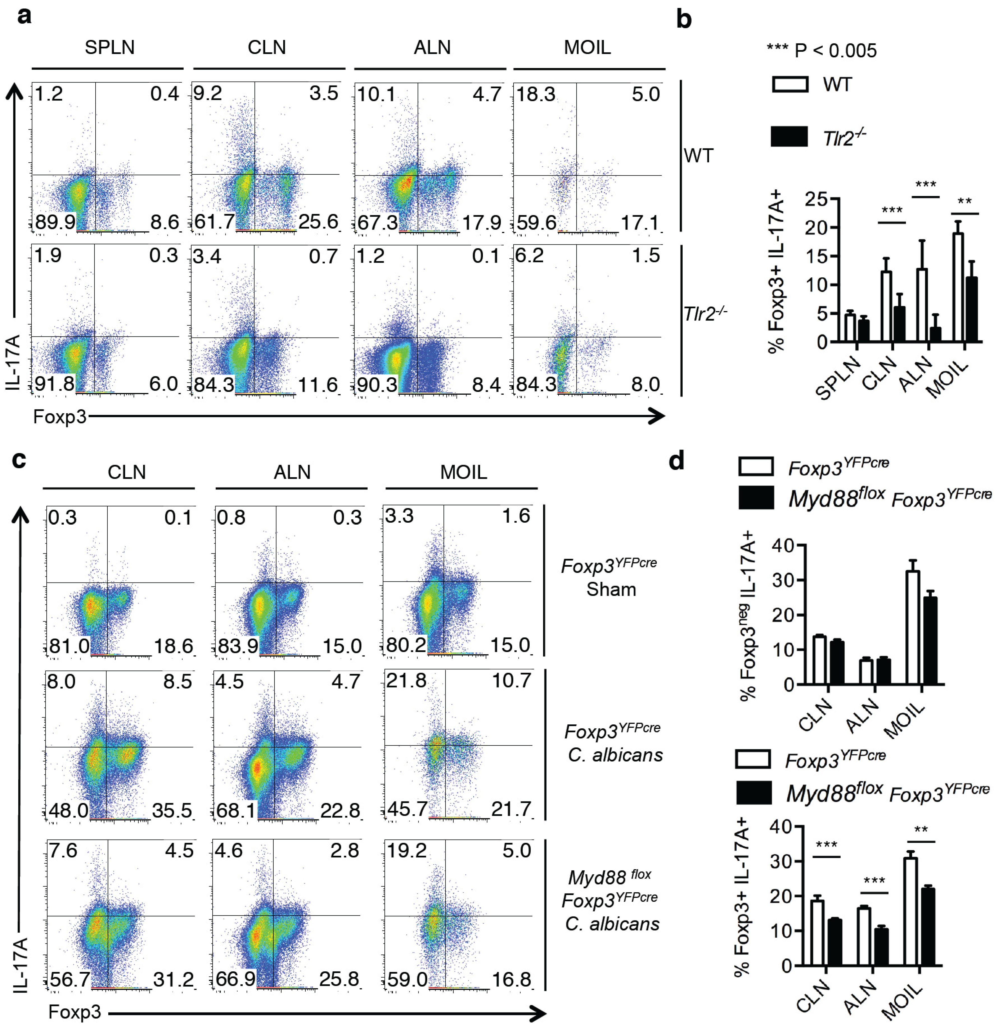
2.6. Direct TLR-2 Signaling in Tregs is Required for Maximal Induction of Foxp3+ IL-17A+ Cells during OPC in Vivo

Lastly, we examined the requirement of TLR-2 expression for inducing IL-17A in Tregs during OPC in vivo. Although TLR-2 signaling promotes Treg proliferation during systemic candidiasis, IL-17A induction was not assessed in Tregs during the infection [21]. To determine the role of TLR-2 signaling in induction of IL-17A in Foxp3+Tregs in vivo, we orally infected WT and Tlr-2−/− mice and measured IL-17A production in CD4+ T cells. We found reduced frequency of Tregs in Tlr-2−/− mice compared to WT mice infiltrating the draining lymphnodes and MOIL during infection (Figure 7a). Importantly, among those Foxp3+CD4+ Tregs the frequency of IL-17A producers was at least three to five times decreased in Tlr-2−/− mice than in WT mice (Figure 7a,b). These data show that TLR-2 signaling enhances proliferation and maximal IL-17A induction in Tregs. Consistent to the role of TLR-2 signaling in promoting IL-17A in effector CD4 cells [14], we also found reduced IL-17A production among Foxp3-effector CD4+ cells in Tlr-2−/− mice, when compared to the WT mice (Figure 7a). To further confirm the direct role of TLR signaling in Tregs, we used Myd88fl°x Foxp3YFPcre mice, in which Myd88 gene was conditionally deleted in Foxp3+ cells. Compared to the control Foxp3YFPcre infected mice, the induction of IL-17A was significantly reduced in Myd88fl°x Foxp3YFPcre mice in Foxp3+ cells (Figure 7c,d lower panel). Although there was a slight decrease in IL-17A production even in the effector cells in MOIL, the frequency of IL-17A producing effector cells was unchanged between the groups in CLN and ALN (Figure 7c,d upper panel). Taken together, these results reveal that direct TLR-2 signaling in Tregs plays a crucial role in increasing IL-17A+Foxp3+ Tregs during OPC in vivo.
Figure 6. IL-17A production of Tregs in an inflammatory milieu is dependent on TLR-2 expression in Tregs and APC. (a,b) WT or Myd88−/− Tregs were stimulated with WT APC or Myd88−/− APC as indicated, without or with Pam3CSK4 or fixed Candida albicans (FCA) germ tube for four days. Foxp3 and IL-17A expression (a), and statistical representation of IL-17A expression in Foxp3+ Tregs (b). α-IL-6 antibodies were added under Th17 conditions without exogenous IL-6 (a, bottom panel). (c,d) WT or Tlr-2−/− Tregs were stimulated with Tlr-2−/− APC, without or with Pam3CSK4 or FCA for 4 days. Foxp3 and IL-17A expression in CD4 gated cells (c), and statistical representation of IL-17A expression (d), are shown. Data represent at least 3 independent experiments.
Pathogens 04 00090 g006
Figure 7. IL-17A expression in CD4+ Foxp3+ Tregs requires TLR-2 signaling in Tregs during C. albicans infection in vivo. WT C57BL/6 or Tlr-2−/− mice infected with C. albicans, and tissues were isolated as in “1a”. Flow cytometric plots gated on CD4+ cells showing IL-17A expression (a), and statistical analyses of IL-17A expression in CD4+Foxp3+ cells (b) are shown. Data represent triplicate experiments. (c,d) Foxp3YFPcre or Myd88 fl°x Foxp3YFPcre mice were infected, and cells were isolated as in “1a”. Flow cytometric plots gated on or CD4+ cells showing IL-17A expression (c), and statistical analyses of IL-17A expression in CD4+Foxp3-cells (d, upper panel) and CD4+Foxp3+ cells (d, lower panel) are shown.
Pathogens 04 00090 g007

3. Discussion

Foxp3+Tregs produce little or no IL-17A in a sterile environment such as thymus in humans or in naïve mice raised under specific pathogen free conditions [6]. Voo et al., were one of the first groups to show the enrichment of peripherally generated IL-17A+Tregs in tonsils, compared to blood, in individuals undergoing tonsillectomy [6]. As tonsils constitute an oral mucosal environment, we hypothesized that oral infections may lead to the generation of these IL-17A+Foxp3+ cells. To verify the tenet, we examined whether they could be induced during oropharyngeal candidiasis infection in mice. Our study shows that OPC can induce Foxp3+IL-17A+Tregs (Treg-17) in a TLR-2/Myd88 dependent manner, revealing one of the mechanisms by which Treg-17 cells are induced during inflammation. We also show that while TLR-2 ligands promote only proliferation in the context of non-inflammatory homeostatic conditions, they can increase proliferation and IL-17A production in Tregs in the context of APC and inflammatory conditions. Although some of these data are in line with the reports that showed TLR-2 signaling induces survival and proliferation in Tregs activated by TCR [12,17], our study also unveils a novel function of TLR-2 in IL-2 dependent homeostatic proliferation of Tregs. Reduced tTregs and pTregs in Tlr-2−/− mice demonstrate that TLR-2 ligands from commensals play a role in maintaining their steady state proliferation (Figure 3b,d). Although TLR-2 signaling was not examined, defective Tregs in germfree and antibiotic treated mice in previous studies [36,37,38,39,40] further confirm the role of commensals in maintaining Tregs in the gut. Further strengthening the idea that TLR-2 signaling induces proliferation of Tregs in vivo, and similar to a previous study [21], we observed reduced Tregs at the site of infection in C. albicans infected Tlr-2−/− mice compared to WT mice. CD4 effector cells and Tregs showed higher TLR-2 protein expression in Th17 inflammatory conditions, compared to Th0 cultures (Figure 5a), showing that Th17 inflammatory cytokines may promote enhanced TLR-2 signaling and proliferation in CD4+T cells [14]. However, contrary to previous findings [15], TLR-2 ligands did not lead to loss of Foxp3 expression. Although under Th17 inflammatory milieu, Tregs did not suppress CD4+ cells, and lost Foxp3 transiently [22], Tregs that expanded with TLR-2 agonists were capable of suppressing fresh CD4+ T cells, upon IL-6 withdrawal. These data imply that the reversal of suppression by TLR-2 agonists in previous studies is likely due to the cytokines in effector CD4 T cells and APC induced by TLR-2 ligands, but not due to their direct effect on Tregs [15]. Apparent loss of Foxp3 expression could also be due to the usage of CD4+CD25+ cells in some studies, instead of CD4+CD25+Foxp3+ cells, that might have led to proliferation of Foxp3 cells in their cultures.
Although TLR-2 ligands can directly induce proliferation of Treg-17 cells, maximal IL-17A induction in Tregs requires IL-6, and TLR-2/Myd88 signaling in APC as well. Mechanistically, although it is conceivable that IL-6 may prime IL-17A induction in Foxp3+Tregs [6], we report here for the first time the role of TLR-2 signaling in increasing the frequency of Treg-17 cells, by promoting proliferation of IL-17A producing Foxp3+Tregs in vitro and in vivo. However, the lower levels of ROR-γt and IL-17A that we observed in Tregs when compared to the effector cells, is likely due to the inhibitory effect of Foxp3 on ROR-γt expression [41,42]. In the context of Candida albicans, even in the absence of TLR-2 signaling in both Tregs and APC, FCA could partially induce IL-17A in Tregs. We speculate that dectin-1 or 2 signaling is also involved in the induction of IL-17A by C. albicans [43]. However, our results showing diminished production of IL-17A in effector CD4+ cells and Tregs in Tlr-2−/− mice, underscore the importance of Candida derived TLR-2 ligands, in orchestrating effector functions and induction of Treg-17 cells during OPC. Treg-17 cells that we describe herein can be induced both from a fraction of tTregs and iTreg cells induced in cultures (Figure 4d). Similarly, Treg-17 cells are possibly derived from tTregs and pTregs during OPC in vivo as well. Whether these Treg-17 cells also play roles in direct antifungal Th17 responses remain to be investigated. This tenet would be consistent with the requirement of Tregs in protective immunity to fungi [24]. However, in the context of suppressing inflammation, Foxp3+ Tregs undergo effector specific functional adaptation in order to suppress distinct effector responses[44]. For example, Tregs acquire effector Tfh characteristics such as Bcl6 and CXCR5 expression to regulate Tfh and germinal center responses in a Bcl-6 dependent manner [45,46]. Likewise, they require IRF-4 and STAT-3 signaling to suppress Th2 and Th17 immunopathology, respectively, in vivo [44,47]. However, it is unknown if Tregs with active STAT-3 signaling express IL-17A to adapt to their environment and engage distinct effector response—specific suppression of Th17 immunopathology. If this is the case, Treg-17 cells promoted by TLR-2 ligands and Th17 cytokines may be a specific population of Tregs programed for antimicrobial functions at early time-points, and for controlling Th17 pathology at later phases of infection. Supporting this notion, the Treg population, which included the effector Treg-17 cells, while enhancing IL-17A in Th17 cells initially, was capable of suppressing OPC immunopathology and IBD immunopathology at later time points in our studies ([22], Figure S1b). Also in vitro, despite acquiring effector functions, >90% of CD25+Foxp3+ Tregs had stable Foxp3 expression, and retained their suppressive properties, upon IL-6 and TLR-2 ligand withdrawal (Figure 4e). Taken together, these data provide important insights into the role of TLR-2 signaling in promoting Treg-17 populations without affecting their suppressive properties during inflammation and infections. We believe that during mucosal inflammation, TLR-2 ligands from commensals and pathogens may drive effector cytokine production in Tregs. Although in the context of local IL-6, Tregs lose their suppressive functions and produce effector cytokines transiently, TLR-2 ligands do not lead to their terminal differentiation in to effector cells. Future studies are warranted to further examine the precise functions of Treg-17 cells during infections and homeostasis in mucosal environments in human diseases.

4. Materials and Methods

4.1. Mice

C57BL/6, Balb/C, C57BL/6 WT, Tlr-2−/−, Il17a−/−cre and Myd88−/−, Foxp3GFP reporter mice, Foxp3YFPcre reporter mice and Myd88fl°x mice were purchased from Jackson Laboratories and were used at 8–12 weeks of age. Myd88fl°x Foxp3YFPcre mice were generated by breeding Foxp3YFPcre reporter mice and Myd88fl°x mice, and Foxp3YFPcre littermates were used as controls. All the mice were maintained in the Animal Resource Center at Case Western Reserve University and cared for in accordance with institutional guidelines and approved IACUC protocols.

4.2. Reagents and Antibodies

Purified α-CD3 (145-2C11), purified α-CD28, α-CD25 (3C7), Biotin α-CD25, fluorescein isothiocyanate (FITC)-α-CD4, phycoerythrin (PE)-α-CD25, unconjugated, (allophycocyanin) APC, Alexafluor-647 or PE conjugated α-CD44, APC, Alexafluor 647, PE or PE-Cy7 conjugated IL-17-A, Foxp3 antibodies, IL-6 blocking antibodies, α-IL-4 and IFN-γ antibodies, were all purchased from eBiosciences (San Diego, CA). Mouse CD4 isolation kit II was purchased from Miltenyi Biotec (Auburn, CA). EasySep isolation kits were purchased from Stem Cell technologies (Canada). Human IL-2 purchased from NCI, was a kind gift from Mike Lenardo lab (NIAID, NIH). Recombinant mouse IL-6 was purchased from BioBasic Inc (Amherst, NY), and human TGF-β was purchased from R&D Systems (Minneapolis, MN). Mouse cells were cultured in complete RPMI-1640 (Bio-Whittaker) supplemented with 10% FCS, 100 U/mL penicillin, 100 μg/mL streptomycin, 5 mM Glutamax (Invitrogen) and 50 μM β-mercaptoethanol.

4.3. Cell Purification

Splenocytes and lymphnodes were harvested from 8- to 12-week-old mice and pooled. CD4 cells were MACS (magnetic assisted cell sorting) purified by using CD4 isolation kit II (Miltenyi Biotec, Auburn, CA) or sorted using EasySep kits. Purified CD4 cells were FACS (Fluorescence Activated Cell Sorting) sorted for CD4+CD25GFP Tcon cells or CD4+CD25+ GFP+ Treg cells (>99% purity). In some experiments CD25+ Tregs and CD4+CD25 CD44l°w naïve Tcon cells were sorted using EasySep kits, and the purity of Tregs was more than 92%. 6 × 10⁴ FACS sorted Tcons or Tregs were cultured in U-bottom 96 well plates with 2 ng/mL IL-2 only, or also with TLR ligands. Pam3CSK4, heat killed Candida albicans (HKCA), Lipopolysaccharide (LPS), and Flagellin were purchased at Invivogen and were used at following concentrations: LPS, 1 ug/mL; Flagellin, 1μg/mL; Pam3CSK4, 5 μg/mL and HKCA, 106/mL. In some experiments, formaldehyde fixed Candida albicans germtube (FCA) were used at 106/mL.

4.4. Th17 Differentiation

Treg cells were cultured alone in U-bottom 96 well plates, or co-cultured with Tcons in the presence of soluble 1 μg/mL of α-CD3 and 2 μg/mL α-CD28 antibodies under Th17 inducing conditions for 3–4 days. Th17 cells were polarized using recombinant mouse IL-6 (25 ng/mL), recombinant human TGF-β (2 ng/mL), recombinant mouse IL-23 (20 ng/mL), recombinant mouse IL-1β (20 ng/mL), α-IFN-γ (3 μg/mL) and α-IL-4 (3 μg/mL). T cell depleted splenocytes were added as antigen presenting cells (APC) in the cultures. Tcons were derived from congenic CD45.1 mice, and Tregs were derived from CD45.2 B6 Foxp3GFP reporter mice so that Tregs could also be tracked using CD45.2 staining. In some experiments, where α-IL-6 antibody was added, we restimulated day-1 Th17 cells under Th17 inducing conditions without adding exogenous IL-6. For “Th0” conditions, cells were stimulated with α-CD3, α-CD28, APC and IL-2 (10 ng/mL). For flow cytometry analyses of IL-17A staining, we used unstained controls and stained Th0 cells as negative controls, to define the gates and mark IL-17A+ cells.

4.5. Quantitative PCR (q-PCR) Analyses

To measure the relative mRNA levels of TLR-2, Foxp3, ROR-γt and IL-17A, RNA was recovered from Tregs or Tcons using EZ-10 RNA isolation kit (BioBasic). Genomic DNA was removed by DNA Away (Ambion; LifeTechnologies (AM1906)) and cDNA was synthesized from using MuLV reverse transcriptase enzyme (BioBasic) and OligodT primers. qPCR reaction was performed using SYBR Green PCR Kit (BioBasic) in a real time PCR machine (BioRad). The relative levels were normalized to the amount of β-actin cDNA levels. The following primer sequences were used: β-actin: 5'-AGCCATGTACGTAGCCATCC-3', 5'-CTCTCAGCTGTGGTGGTGAA-3', TLR-2: 5'-CCGGAATTATCAGTCCCAAA-3', 5'-ATCTCCAGCAGGAAAGCAGA-3', ROR-γt: 5'-AACAGGAACAAGTGGCCAAG-3', 5'-GGTAGCTGCCCATCTGAGAG-3', FoxP3: 5'-TTCATGCATCAGCTCTCCAC-3', 5'-TGATCATGGCTGGGTTGTC-3', IL-17A: 5'-AAAGCTCAGCGTGTCCAAA-3', 5'-GCGCCAAGGGAGTTAAAGAC-3'.

4.6. C. albicans Infection in Mice

Age and sex matched mice were infected and individually caged after infection, as previously described [22,48]. They were infected under anesthesia by placing a 3 mm diameter cotton ball saturated with PBS (Sham) or 2 × 10⁷ C. albicans (CAF2-1) blastospores sublingually for 90 min. Spleen, ALN, CLN, and MOIL were harvested, one or two days after infection for phorbol myrisate acetate (PMA), ionomycin and brefeldin-A re-stimulation, and intracellular cytokine analyses.

4.7. IBD Induction by Th17 Cell Transfer in Vivo

For Th17 IBD, CD45.1 Rag1−/− mice received 8 × 10⁵ CD45.1 Th17 cells that were stimulated and differentiated for 5 days as above. Two weeks later, the mice received fresh CD45.2 Tregs derived from congenic CD45.2 Foxp3GFP reporter mice. Spleen, MLN and the gut LP cells were harvested 5 days after Treg injection, for PMA/ionomycin/brefeldin-A re-stimulation and intracellular cytokine analyses.

4.8. Statistical Analyses

p values were calculated by Students “t” test in Microsoft Excel software using two tailed distribution and two-sample equal variance parameters, with ** being p < 0.05. We also used Mann-Whitney tests, or 2 way ANOVA Sidak’s multiple comparisons test (alpha value 0.05; 95% CI) in Prism 6.0 (GraphPad Software, Inc.) to calculate significance.

5. Conclusions

CD4+Foxp3+Tregs produce the effector cytokine IL-17A in vitro and in vivo during oropharyngeal candidiasis (OPC) in a TLR-2/Myd88 signaling dependent manner. We call these cells as Treg-17 cells. Although TLR-2/Myd88 signaling can directly induce proliferation of Treg-17 cells, maximal IL-17A induction in Tregs requires IL-6, and TLR-2 signaling in APC as well. The minimal and transient loss of Foxp3 expression and suppressive properties are due to the presence of IL-6 in the milieu, but not the direct effect of TLR-2 signaling in Tregs. Taken together, our data reveal that TLR-2 signaling promotes not only proliferation, but also IL-17A in Tregs, depending on the cytokine milieu. These Treg-17 cells may be relevant in mucosal infections and inflammation.

Supplementary Materials

Supplementary materials can be accessed at: https://www.mdpi.com/2076-0817/4/1/90/s1.

Acknowledgments

The project is supported by the startup funding to PP from the Department of Biological Sciences, SODM, Case Western Reserve University and skin disease research center (SDRC) grants to PP. We also thank Center For AIDS Research (CFAR) immune function core for access to BD Fortessa flow cytometer.

Authors Contributions

PP conceived, designed, and performed replicate experiments; acquired, analyzed and interpreted the data; drafted and revised the manuscript. NB and YZ were involved in performing the cell isolation procedures and performing cell culture experiments. SC performed qPCR experiments and analyzed the data. AW critically reviewed and revised the manuscript critically.

Conflicts of Interest

The authors declare no conflict of interest.

References

  1. Pandiyan, P.; Zheng, L.; Lenardo, M.J. The molecular mechanisms of regulatory T cell immunosuppression. Front. Immunol. 2011, 2, 60. [Google Scholar] [CrossRef] [PubMed]
  2. Rudensky, A.Y.; Campbell, D.J. In vivo sites and cellular mechanisms of t reg cell-mediated suppression. J. Exp. Med. 2006, 203, 489–492. [Google Scholar] [CrossRef] [PubMed]
  3. Beriou, G.; Costantino, C.M.; Ashley, C.W.; Yang, L.; Kuchroo, V.K.; Baecher-Allan, C.; Hafler, D.A. Il-17-producing human peripheral regulatory T cells retain suppressive function. Blood 2009, 113, 4240–4249. [Google Scholar] [CrossRef] [PubMed]
  4. Du, R.; Zhao, H.; Yan, F.; Li, H. Il-17+foxp3+ T cells: An intermediate differentiation stage between th17 cells and regulatory T cells. J. Leukoc. Biol. 2014, 96, 39–48. [Google Scholar] [CrossRef] [PubMed]
  5. Hovhannisyan, Z.; Treatman, J.; Littman, D.R.; Mayer, L. Characterization of interleukin-17-producing regulatory T cells in inflamed intestinal mucosa from patients with inflammatory bowel diseases. Gastroenterology 2011, 140, 957–965. [Google Scholar] [CrossRef] [PubMed]
  6. Voo, K.S.; Wang, Y.H.; Santori, F.R.; Boggiano, C.; Wang, Y.H.; Arima, K.; Bover, L.; Hanabuchi, S.; Khalili, J.; Marinova, E.; et al. Identification of il-17-producing foxp3+ regulatory T cells in humans. Proc. Natl. Acad. Sci. USA 2009, 106, 4793–4798. [Google Scholar] [CrossRef] [PubMed]
  7. Whibley, N.; Maccallum, D.M.; Vickers, M.A.; Zafreen, S.; Waldmann, H.; Hori, S.; Gaffen, S.L.; Gow, N.A.; Barker, R.N.; Hall, A.M. Expansion of foxp3(+) t-cell populations by candida albicans enhances both th17-cell responses and fungal dissemination after intravenous challenge. Eur. J. Immunol. 2014, 44, 1069–1083. [Google Scholar] [CrossRef] [PubMed] [Green Version]
  8. Colonna, L.; Florek, M.; Leveson-Gower, D.B.; Sega, E.I.; Baker, J.; Smith, A.T.; Negrin, R.S. Il-17 gene ablation does not impact treg-mediated suppression of graft-versus-host disease after bone marrow transplantation. Biol. Blood Marrow Transplant. 2013, 19, 1557–1565. [Google Scholar] [CrossRef] [PubMed]
  9. Takeuchi, O.; Sato, S.; Horiuchi, T.; Hoshino, K.; Takeda, K.; Dong, Z.; Modlin, R.L.; Akira, S. Cutting edge: Role of toll-like receptor 1 in mediating immune response to microbial lipoproteins. J. Immunol. 2002, 169, 10–14. [Google Scholar] [CrossRef] [PubMed]
  10. Ishii, K.J.; Koyama, S.; Nakagawa, A.; Coban, C.; Akira, S. Host innate immune receptors and beyond: Making sense of microbial infections. Cell Host Microbe 2008, 3, 352–363. [Google Scholar] [CrossRef] [PubMed]
  11. Uematsu, S.; Akira, S. Toll-like receptors (tlrs) and their ligands. Handb. Exp. Pharmacol. 2008, 183, 1–20. [Google Scholar] [PubMed]
  12. Sutmuller, R.P.; Morgan, M.E.; Netea, M.G.; Grauer, O.; Adema, G.J. Toll-like receptors on regulatory T cells: Expanding immune regulation. Trends Immunol. 2006, 27, 387–393. [Google Scholar] [CrossRef] [PubMed]
  13. Liu, J.; Radler, D.; Illi, S.; Klucker, E.; Turan, E.; von Mutius, E.; Kabesch, M.; Schaub, B. Tlr2 polymorphisms influence neonatal regulatory T cells depending on maternal atopy. Allergy 2011, 66, 1020–1029. [Google Scholar] [CrossRef] [PubMed]
  14. Reynolds, J.M.; Pappu, B.P.; Peng, J.; Martinez, G.J.; Zhang, Y.; Chung, Y.; Ma, L.; Yang, X.O.; Nurieva, R.I.; Tian, Q.; et al. Toll-like receptor 2 signaling in cd4(+) t lymphocytes promotes T helper 17 responses and regulates the pathogenesis of autoimmune disease. Immunity 2010, 32, 692–702. [Google Scholar] [CrossRef] [PubMed]
  15. Liu, H.; Komai-Koma, M.; Xu, D.; Liew, F.Y. Toll-like receptor 2 signaling modulates the functions of cd4+ cd25+ regulatory T cells. Proc. Natl. Acad. Sci. USA 2006, 103, 7048–7053. [Google Scholar] [CrossRef] [PubMed]
  16. Van Maren, W.W.; Nierkens, S.; Toonen, L.W.; Bolscher, J.M.; Sutmuller, R.P.; Adema, G.J. Multifaceted effects of synthetic tlr2 ligand and legionella pneumophilia on treg-mediated suppression of T cell activation. BMC Immunol. 2011, 12, 23. [Google Scholar]
  17. Chen, Q.; Davidson, T.S.; Huter, E.N.; Shevach, E.M. Engagement of tlr2 does not reverse the suppressor function of mouse regulatory T cells, but promotes their survival. J. Immunol. 2009, 183, 4458–4466. [Google Scholar] [CrossRef] [PubMed]
  18. Round, J.L.; Lee, S.M.; Li, J.; Tran, G.; Jabri, B.; Chatila, T.A.; Mazmanian, S.K. The toll-like receptor 2 pathway establishes colonization by a commensal of the human microbiota. Science 2011, 332, 974–977. [Google Scholar] [CrossRef] [PubMed]
  19. Pasare, C.; Medzhitov, R. Toll pathway-dependent blockade of cd4+cd25+ T cell-mediated suppression by dendritic cells. Science 2003, 299, 1033–1036. [Google Scholar] [CrossRef] [PubMed]
  20. Kubo, T.; Hatton, R.D.; Oliver, J.; Liu, X.; Elson, C.O.; Weaver, C.T. Regulatory T cell suppression and anergy are differentially regulated by proinflammatory cytokines produced by tlr-activated dendritic cells. J. Immunol. 2004, 173, 7249–7258. [Google Scholar] [CrossRef] [PubMed]
  21. Netea, M.G.; Sutmuller, R.; Hermann, C.; van der Graaf, C.A.; van der Meer, J.W.; van Krieken, J.H.; Hartung, T.; Adema, G.; Kullberg, B.J. Toll-like receptor 2 suppresses immunity against candida albicans through induction of il-10 and regulatory T cells. J. Immunol. 2004, 172, 3712–3718. [Google Scholar] [CrossRef] [PubMed]
  22. Pandiyan, P.; Conti, H.R.; Zheng, L.; Peterson, A.C.; Mathern, D.R.; Hernandez-Santos, N.; Edgerton, M.; Gaffen, S.L.; Lenardo, M.J. Cd4(+)cd25(+)foxp3(+) regulatory T cells promote th17 cells in vitro and enhance host resistance in mouse candida albicans th17 cell infection model. Immunity 2011, 34, 422–434. [Google Scholar] [CrossRef] [PubMed]
  23. Whibley, N.; Gaffen, S.L. Brothers in arms: Th17 and treg responses in candida albicans immunity. PLoS Pathog. 2014, 10, e1004456. [Google Scholar] [CrossRef] [PubMed]
  24. Montagnoli, C.; Bacci, A.; Bozza, S.; Gaziano, R.; Mosci, P.; Sharpe, A.H.; Romani, L. B7/cd28-dependent cd4+cd25+ regulatory T cells are essential components of the memory-protective immunity to candida albicans. J. Immunol. 2002, 169, 6298–6308. [Google Scholar] [CrossRef] [PubMed]
  25. Hernandez-Santos, N.; Huppler, A.R.; Peterson, A.C.; Khader, S.A.; McKenna, K.C.; Gaffen, S.L. Th17 cells confer long-term adaptive immunity to oral mucosal candida albicans infections. Mucosal Immunol. 2013, 6, 900–910. [Google Scholar] [CrossRef] [PubMed]
  26. Pandiyan, P.; Bhaskaran, N.; Zhang, Y.; Weinberg, A. Isolation of T cells from mouse oral tissues. Biol. Proced. Online 2014, 16, 4. [Google Scholar] [CrossRef] [PubMed]
  27. Izcue, A.; Coombes, J.L.; Powrie, F. Regulatory lymphocytes and intestinal inflammation. Annu. Rev. Immunol. 2009, 27, 313–338. [Google Scholar] [CrossRef] [PubMed]
  28. Gil, M.L.; Gozalbo, D. Role of toll-like receptors in systemic candida albicans infections. Front. Biosci. 2009, 14, 570–582. [Google Scholar] [CrossRef]
  29. Gratz, I.K.; Rosenblum, M.D.; Abbas, A.K. The life of regulatory T cells. Ann. NY Acad. Sci. 2013, 1283, 8–12. [Google Scholar] [CrossRef] [PubMed]
  30. Pandiyan, P.; Lenardo, M.J. The control of cd4+cd25+foxp3+ regulatory T cell survival. Biol. Direct 2008, 3, 6. [Google Scholar] [CrossRef] [PubMed]
  31. Turka, L.A.; Walsh, P.T. Il-2 signaling and cd4+ cd25+ foxp3+ regulatory T cells. Front. Biosci. 2008, 13, 1440–1446. [Google Scholar] [CrossRef] [PubMed]
  32. Xu, L.; Kitani, A.; Fuss, I.; Strober, W. Cutting edge: Regulatory T cells induce cd4+cd25-foxp3-t cells or are self-induced to become th17 cells in the absence of exogenous tgf-beta. J. Immunol. 2007, 178, 6725–6729. [Google Scholar] [CrossRef] [PubMed]
  33. Lal, G.; Zhang, N.; van der Touw, W.; Ding, Y.; Ju, W.; Bottinger, E.P.; Reid, S.P.; Levy, D.E.; Bromberg, J.S. Epigenetic regulation of foxp3 expression in regulatory T cells by DNA methylation. J. Immunol. 2009, 182, 259–273. [Google Scholar] [CrossRef] [PubMed]
  34. Pandiyan, P.; Zheng, L.; Ishihara, S.; Reed, J.; Lenardo, M.J. Cd4(+)cd25(+)foxp3(+) regulatory T cells induce cytokine deprivation-mediated apoptosis of effector cd4(+) T cells. Nat. Immunol. 2007, 8, 1353–1362. [Google Scholar] [CrossRef] [PubMed]
  35. Takeuchi, O.; Akira, S. Myd88 as a bottle neck in toll/il-1 signaling. Curr. Top. Microbiol. Immunol. 2002, 270, 155–167. [Google Scholar] [PubMed]
  36. Atarashi, K.; Tanoue, T.; Shima, T.; Imaoka, A.; Kuwahara, T.; Momose, Y.; Cheng, G.; Yamasaki, S.; Saito, T.; Ohba, Y.; et al. Induction of colonic regulatory T cells by indigenous clostridium species. Science 2011, 331, 337–341. [Google Scholar] [CrossRef] [PubMed]
  37. Cording, S.; Wahl, B.; Kulkarni, D.; Chopra, H.; Pezoldt, J.; Buettner, M.; Dummer, A.; Hadis, U.; Heimesaat, M.; Bereswill, S.; et al. The intestinal micro-environment imprints stromal cells to promote efficient treg induction in gut-draining lymph nodes. Mucosal. Immunol. 2013, 7, 359–368. [Google Scholar] [CrossRef] [PubMed]
  38. Ostman, S.; Rask, C.; Wold, A.E.; Hultkrantz, S.; Telemo, E. Impaired regulatory T cell function in germ-free mice. Eur. J. Immunol. 2006, 36, 2336–2346. [Google Scholar] [CrossRef] [PubMed]
  39. Smith, P.M.; Howitt, M.R.; Panikov, N.; Michaud, M.; Gallini, C.A.; Bohlooly, Y.M.; Glickman, J.N.; Garrett, W.S. The microbial metabolites, short-chain fatty acids, regulate colonic treg cell homeostasis. Science 2013, 341, 569–573. [Google Scholar] [CrossRef] [PubMed]
  40. Strauch, U.G.; Obermeier, F.; Grunwald, N.; Gurster, S.; Dunger, N.; Schultz, M.; Griese, D.P.; Mahler, M.; Scholmerich, J.; Rath, H.C. Influence of intestinal bacteria on induction of regulatory T cells: Lessons from a transfer model of colitis. Gut 2005, 54, 1546–1552. [Google Scholar] [CrossRef] [PubMed]
  41. Zeng, W.P.; Chang, C.; Lai, J.J. Immune suppressive activity and lack of t helper differentiation are differentially regulated in natural regulatory T cells. J. Immunol. 2009, 183, 3583–3590. [Google Scholar] [CrossRef] [PubMed]
  42. Zhou, Z.; Song, X.; Li, B.; Greene, M.I. Foxp3 and its partners: Structural and biochemical insights into the regulation of foxp3 activity. Immunologic research 2008, 42, 19–28. [Google Scholar] [CrossRef] [PubMed]
  43. Cheng, S.C.; van de Veerdonk, F.L.; Lenardon, M.; Stoffels, M.; Plantinga, T.; Smeekens, S.; Rizzetto, L.; Mukaremera, L.; Preechasuth, K.; Cavalieri, D.; et al. The dectin-1/inflammasome pathway is responsible for the induction of protective t-helper 17 responses that discriminate between yeasts and hyphae of candida albicans. J. Leukoc. Biol. 2011, 90, 357–366. [Google Scholar] [CrossRef] [PubMed]
  44. Chaudhry, A.; Rudensky, A.Y. Control of inflammation by integration of environmental cues by regulatory T cells. J. Clin. Investig. 2013, 123, 939–944. [Google Scholar] [CrossRef] [PubMed]
  45. Chung, Y.; Tanaka, S.; Chu, F.; Nurieva, R.I.; Martinez, G.J.; Rawal, S.; Wang, Y.H.; Lim, H.; Reynolds, J.M.; Zhou, X.H.; et al. Follicular regulatory T cells expressing foxp3 and bcl-6 suppress germinal center reactions. Nat. Med. 2011, 17, 983–988. [Google Scholar] [CrossRef] [PubMed]
  46. Linterman, M.A.; Pierson, W.; Lee, S.K.; Kallies, A.; Kawamoto, S.; Rayner, T.F.; Srivastava, M.; Divekar, D.P.; Beaton, L.; Hogan, J.J.; et al. Foxp3+ follicular regulatory T cells control the germinal center response. Nat. Med. 2011, 17, 975–982. [Google Scholar] [CrossRef] [PubMed]
  47. Chaudhry, A.; Rudra, D.; Treuting, P.; Samstein, R.M.; Liang, Y.; Kas, A.; Rudensky, A.Y. Cd4+ regulatory T cells control th17 responses in a stat3-dependent manner. Science 2009, 326, 986–991. [Google Scholar] [CrossRef]
  48. Kamai, Y.; Kubota, M.; Kamai, Y.; Hosokawa, T.; Fukuoka, T.; Filler, S.G. New model of oropharyngeal candidiasis in mice. Antimicrob. Agents Chemother. 2001, 45, 3195–3197. [Google Scholar] [CrossRef] [PubMed]

